(주)제이엔옵틱 특허품인 JNO-MHU <Measuring Height unit> 와 단면 관찰이 용이하게 절단해주는 JNO-FM-JIG 장착 모델 입니다.
(주)제이엔옵틱 특허품인 JNO-MHU <Measuring Height unit> 와 단면 관찰이 용이하게 절단해주는 JNO-FM-JIG 장착 모델 입니다.
저배율 쌍안 실체현미경입니다.
외관 검사용으로 적합합니다.
기본 배율 : 6.7배 ~ 45배
작업거리 : 100mm
베이스 : 320 X 260mm
접안렌즈 : 10배 (2ea)
링 타입 LED조명
원산지 : 중국
공급가 : 50만원(VAT 포함)
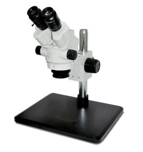
줌 타입 비디오 현미경 SET
줌 기본 배율 0.7배 ~ 4.5배, 대물렌즈 1배, 카메라 아답타0.5배, 스텐드 베이스 370x258x323mm, 25Ø (SZ5D40-S)
LED 조명+ 기본형 스텐드 + ZOOM LENS + 카메라
자세한 사항은 문의 바랍니다. 02-3473-4188
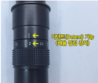
Detent 기능으로 배율 변환시 고정적으로 사용 가능 합니다.
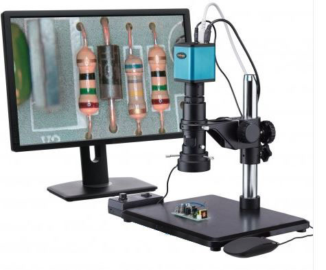
고화질의 카메라와 모니터를 적용하여 검사실 환경과 작업 피로도를 줄여 생산성 향상에 기여 합니다.
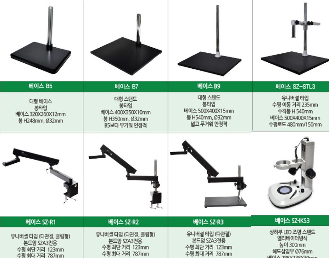
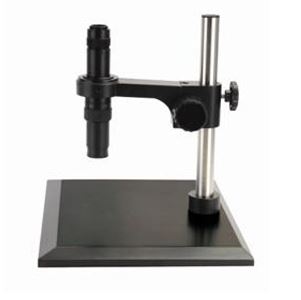
다양한 타입의 스텐드로 변경 가능!!
1배 대물렌즈 장착시 : 7배~ 45배, 초점거리: 96mm
보이는 영역 20mm X 15mm ~ 2.7mm X 2.1mm
줌 현미경과 1/2″카메라 사용시 보이는 영역을 캡쳐한 이미지 입니다.
가장 낮은 배율인 경우 가로 약2cm 세로 약1.5cm 가 보입니다
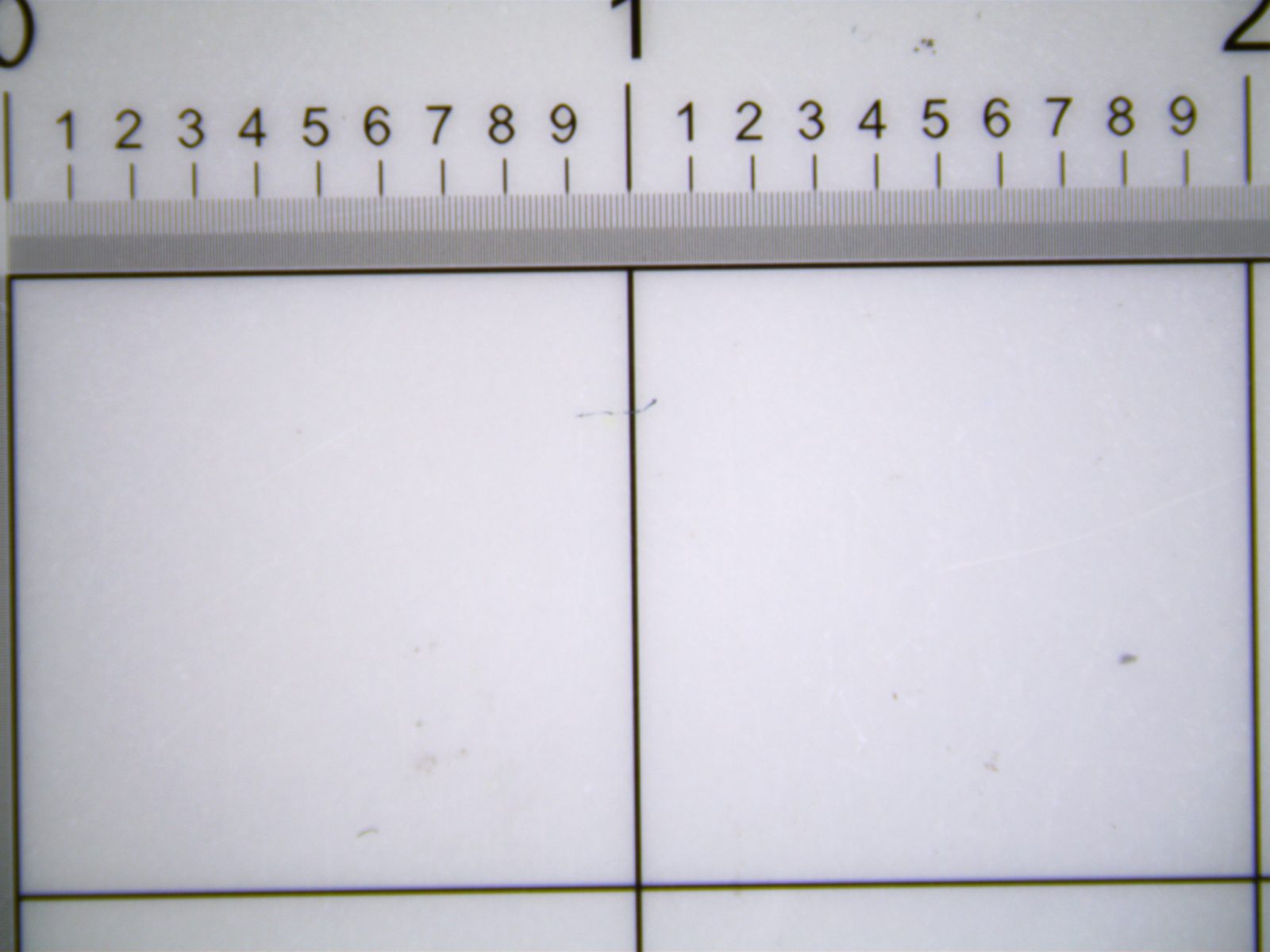
가장 높은 배율인 경우 가로 약2.7mm 세로약2.1mm
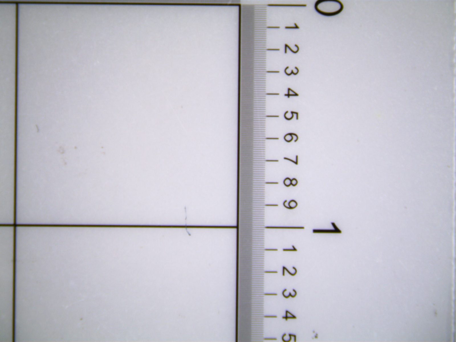
0.5배 대물렌즈 장착시 : 3.5배 ~ 23배, 초점거리 : 198mm
보이는 영역 5.7m X 4.4mm ~ 2.7mm X 2.1mm
줌 현미경과 1/2″카메라 사용시 보이는 영역을 캡쳐한 이미지 입니다.
가장 낮은 배율인 경우 가로 약4.4cm 세로 약3.2cm 가 보입니다.

가장 높은 배율인 경우 가로 약5.7mm 세로약4.4mm
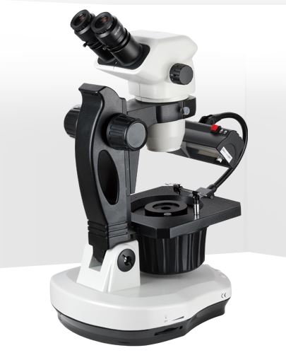
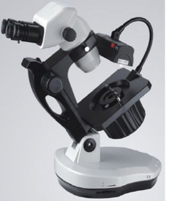
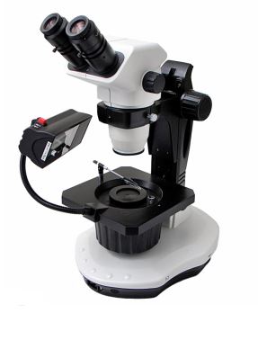
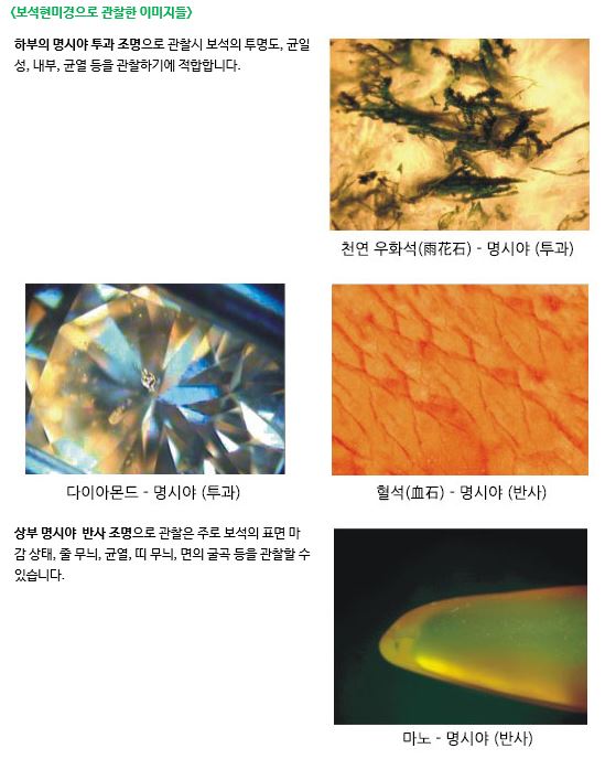
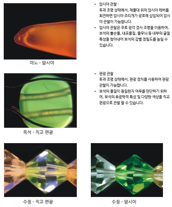
보석관찰에 최적화된 현미경입니다.
쌍안 타입
총 관찰 배율 : 6.7배 ~ 45배
45도 경사의 인체공학적 헤드, 작업거리 100mm
다양한 조명으로 보석 관찰 :
형광 및 할로겐 조명, 평행 /사선/투과/반사 조명,
명시야/암시야(옵션)/편광 조명 관찰
상부 (반사조명) 하부 (투과조명)
보석 관찰에 최적화된 스탠드 :
디스크 360도 회전, 관찰 각도 조절
본체 리프팅 가능
보석 고정이 편리한 젬 클램프 (Gem clamp) 장착
공급가 1,430,000원 암시야관찰시 110,000원 추가 됩니다.
(부가세포함가격 입니다.)
기타 문의 사항은 연락 바랍니다. 02-3473-4188
삼안 줌 실체 현미경 6.7배~45배
10배 접안렌즈 , 105mm 작업거리
고휘도 링타입 LED 조명장치
200x255x 22mm (300mm)상하 조절용 스텐드
300만 화소 디지탈 카메라
현미경 + LED 조명 + 300만 화소 디지탈 카메라(측정 SW포함)
자세한 문의 사항은 02-3473-4188 연락 바랍니다
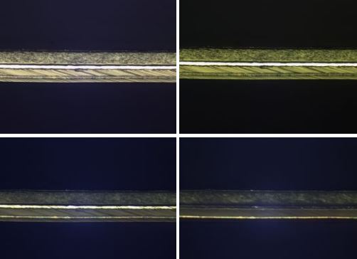
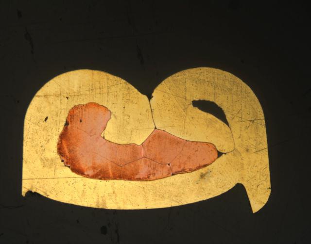
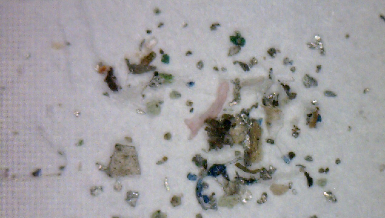
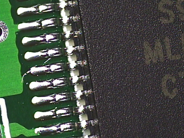
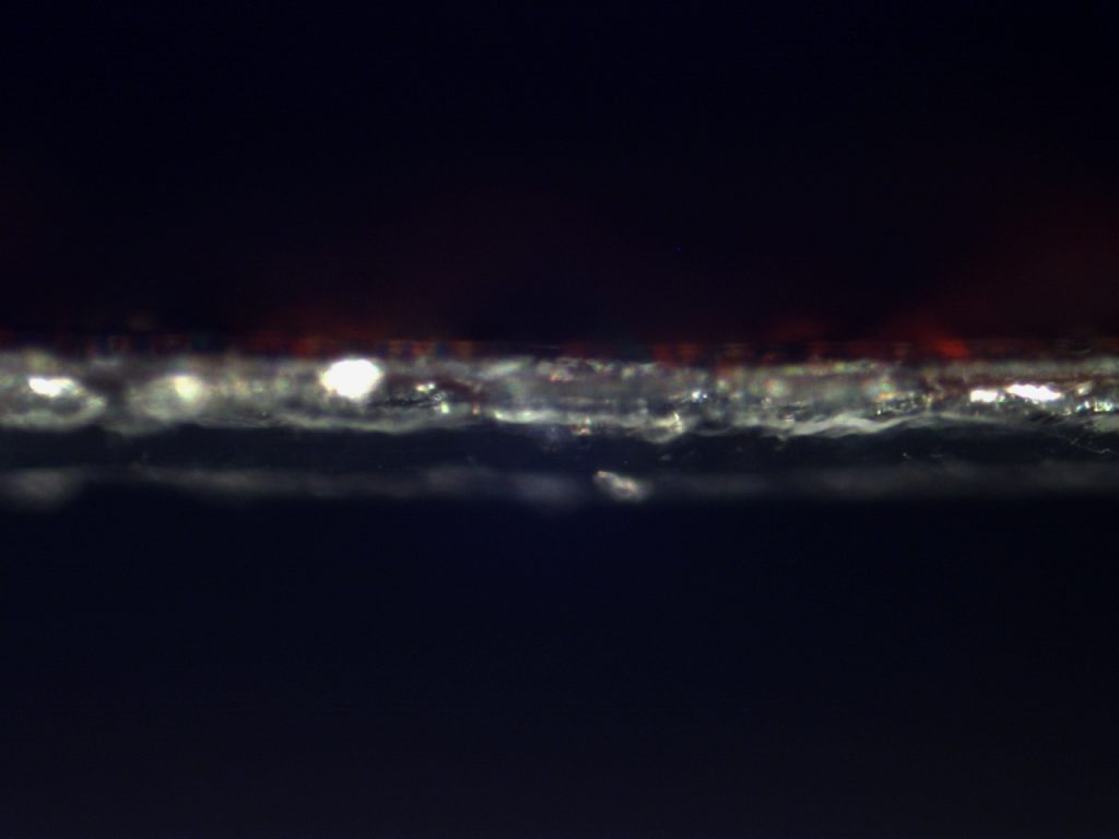
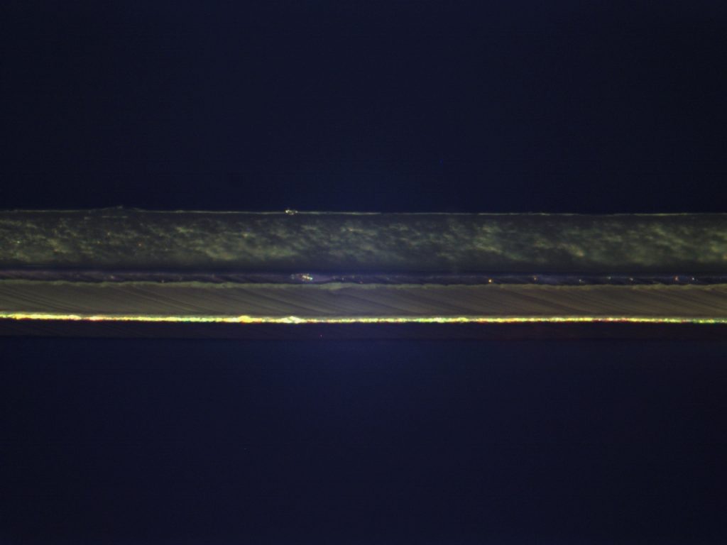
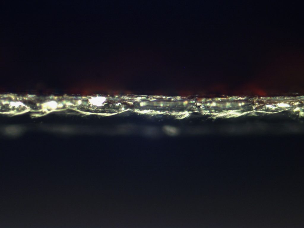
각종 적용 사례
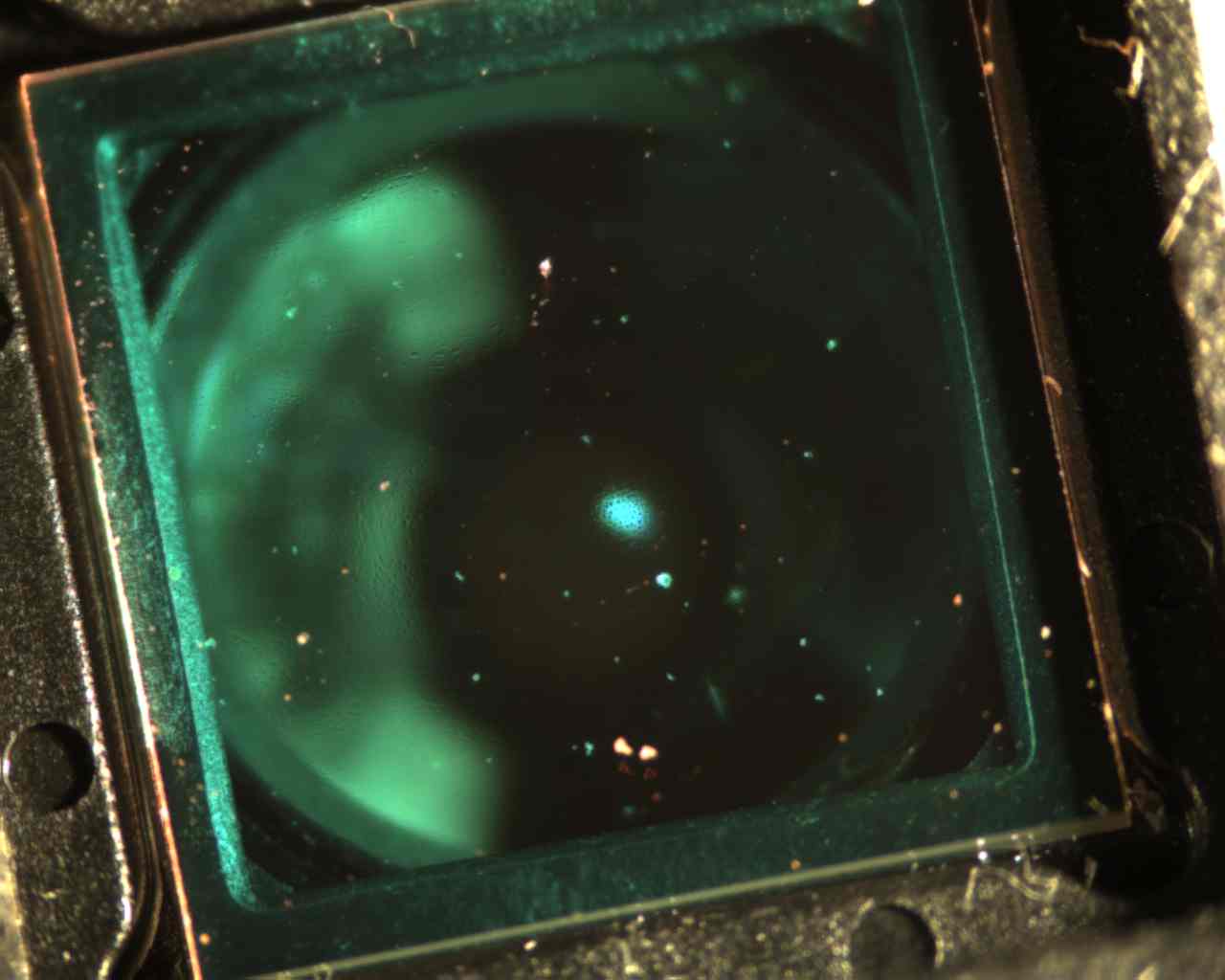
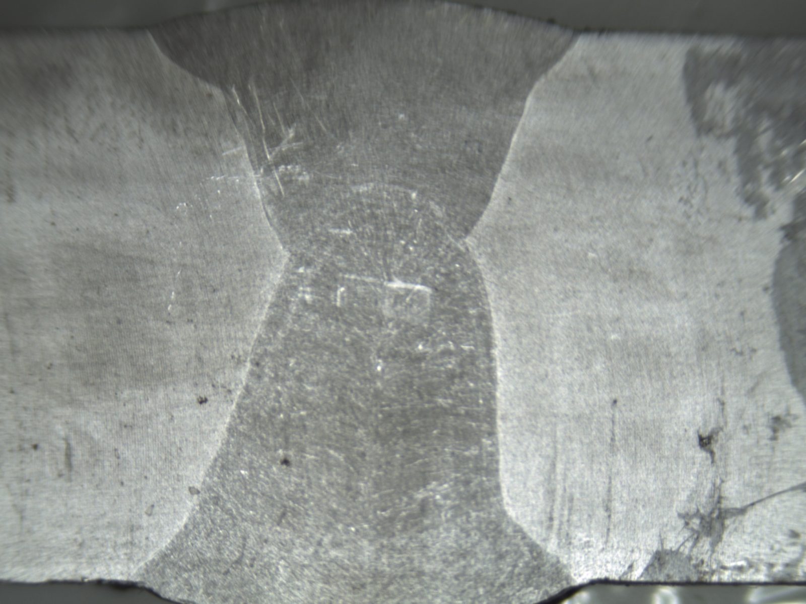
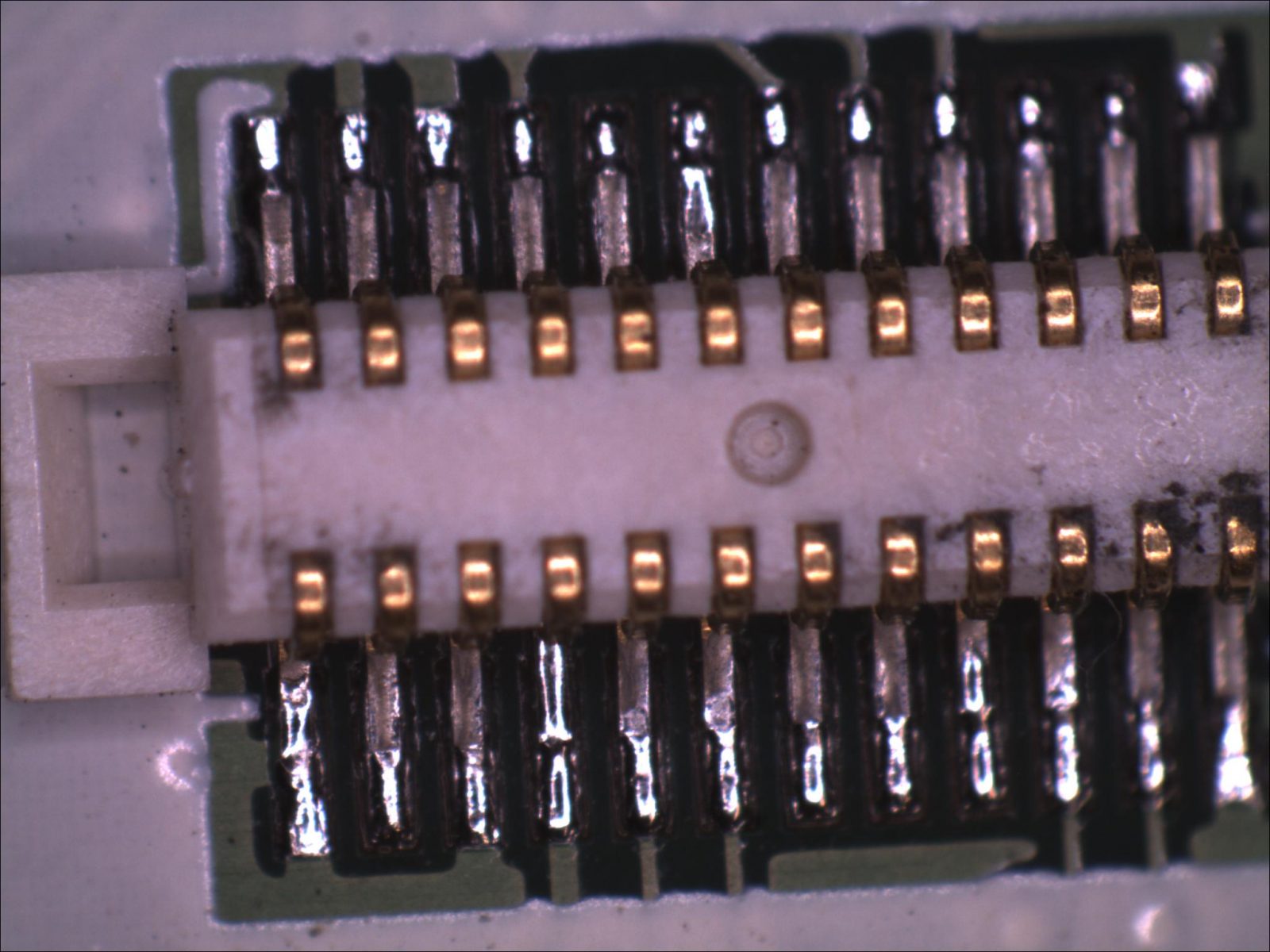
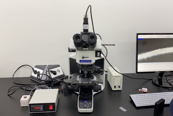
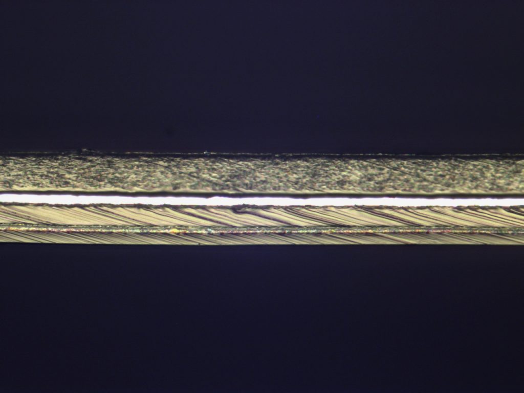
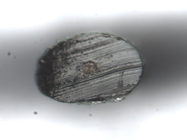
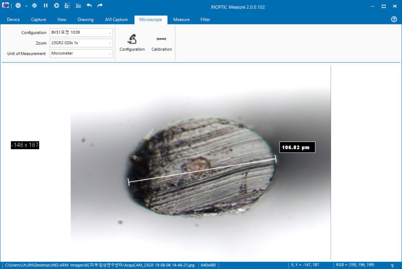
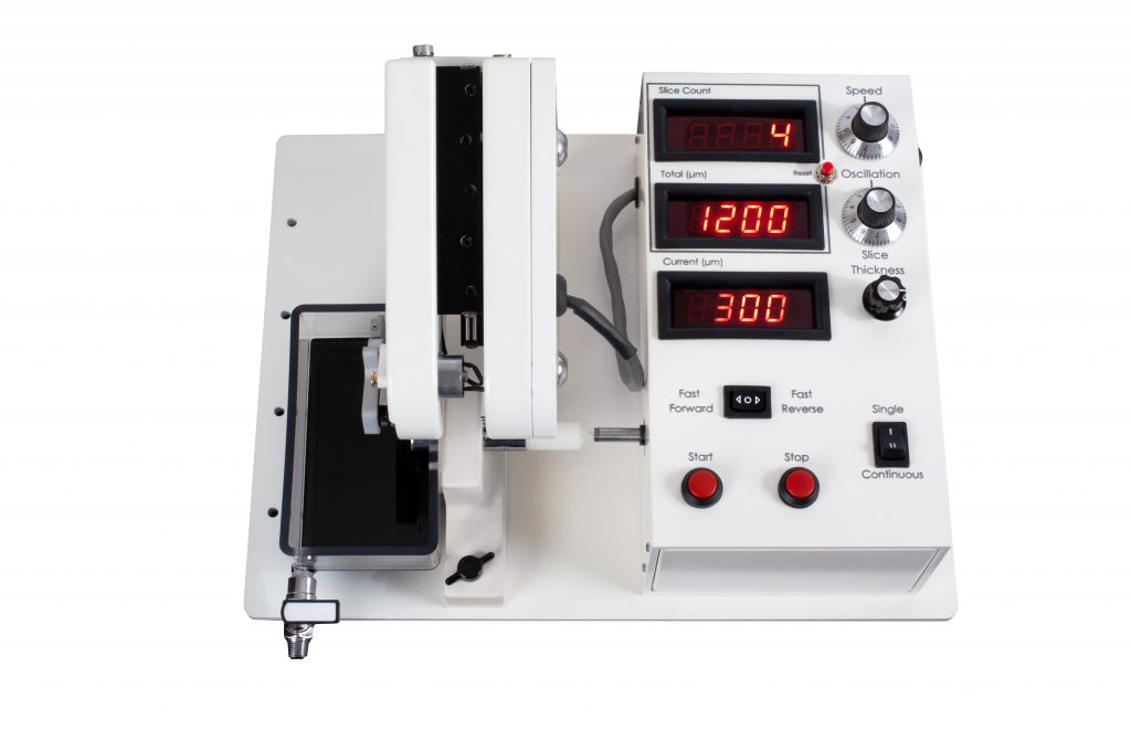
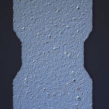
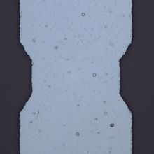
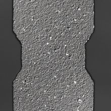
필름 단면 관찰 현미경
JNO-FM-BX53-SET
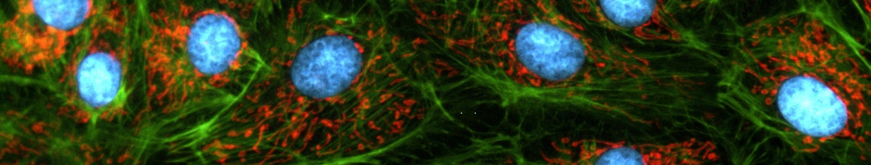
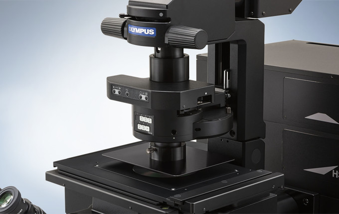
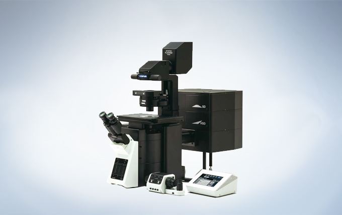
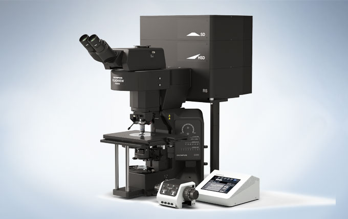
The FLUOVIEW FV3000 series is designed to meet some of the most difficult challenges in modern science. Featuring the high sensitivity and speed required for live cell and tissue imaging, the FV3000 enables 2D-6D (x,y,z,t,λ,p) macro to micro imaging of cells, tissues, and small organisms. With an intuitive and adaptable user interface, the FV3000 supports complete workflows from image acquisition to processing and analysis. Particular attention has been paid to the needs of cell biology, cancer research, and stem cell research, and with two new upright configurations, the FV3000 is also poised to meet the needs of neuroscience, electrophysiology, and developmental biology.
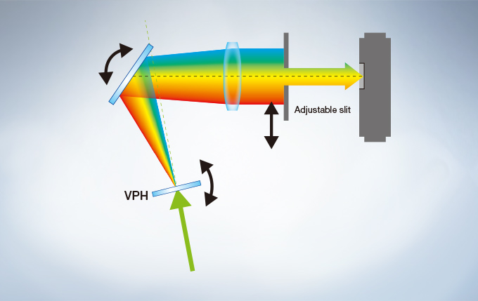
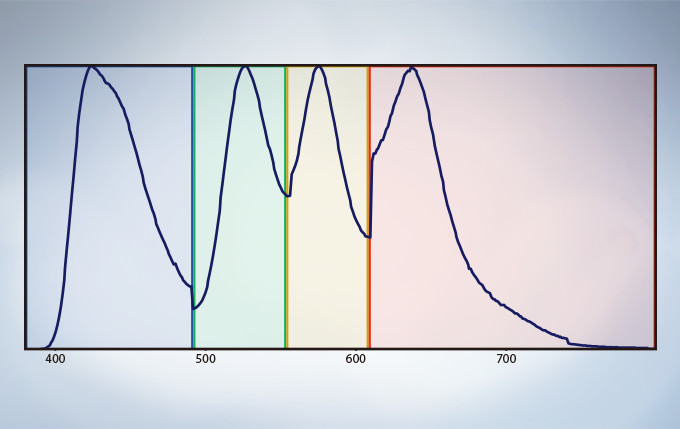
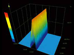
The FV3000 series employs Olympus’ TruSpectral detection technology. Based on patented* Volume Phase Hologram (VPH) transmission and an adjustable slit to control light, the spectral detection is highly efficient, enabling users to select the detection wavelength of each individual channel to 2 nm.
Efficient TruSpectral Detection System
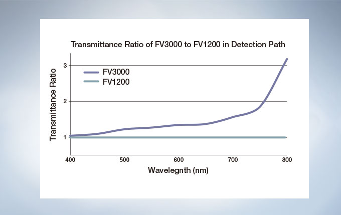
The FV3000 is a fully spectral series of confocal microscope. TruSpectral detection delivers improved overall transmission and sensitivity. The high signal-to-noise ratio results in excellent multi-color confocal imaging capabilities.
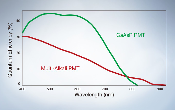
The GaAsP Photomultiplier Tubes (PMTs) in the FV3000’s high sensitivity detector (HSD) enable users to view samples whose emission is too weak to view with conventional detection methods. The GaAsP PMT unit incorporates two channels with a maximum quantum efficiency of 45%, and Peltier cooling that reduces background noise by 20% for high S/N ratio images under very low excitation light.
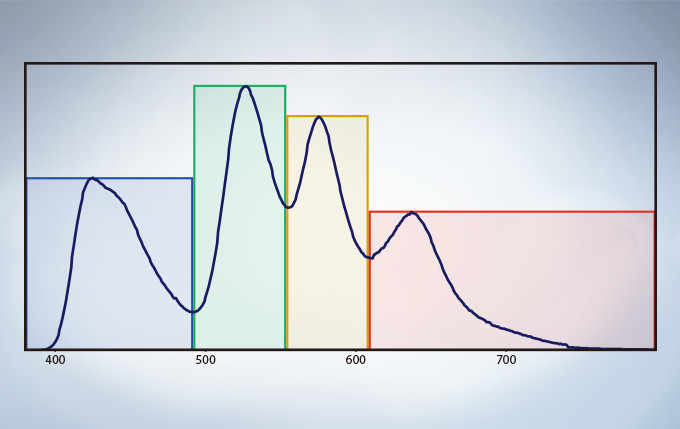
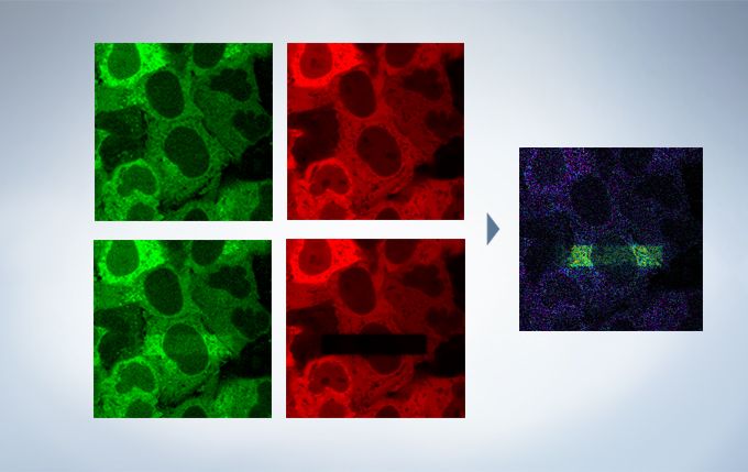
TruSpectral technology’s efficient design and software enable spectral detectors to run in multichannel mode for both live and post-processing spectral unmixing with a multichannel lambda mode. The multichannel mode facilitates constant spectral unmixing during live cell experiments, separating complex fluorescence during acquisition. With up to four different dynamic ranges from the four different channels of array, bright and dim spectral signals can be separated by independently adjusting the sensitivity of each detector.
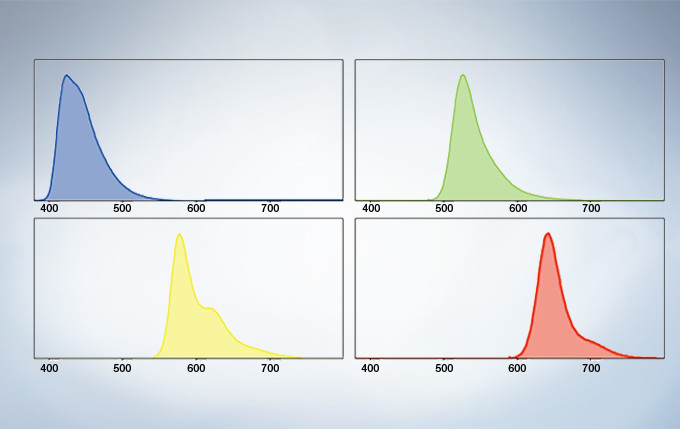
The deconvolution algorithm enables overlapping spectra to be separated based on the spectral information from lambda stack images. The fluorescence cross-talk between the channels can be eliminated by the unmixing algorithm during both image acquisition and post acquisition processing.
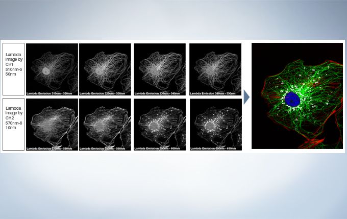
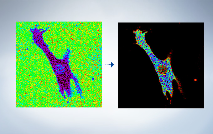
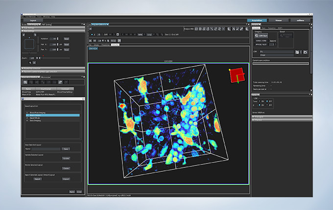
Live Spectral Unmixing with TruSpectral Detection and Real-Time Processing
The power of TruSpectral detection plus multichannel mode means live spectral unmixing can be performed during image acquisition. Complex, overlapping spectra can be processed in real time.
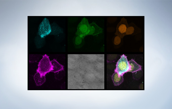
Image data courtesy of Dr. Kazuhiro Aoki, Dr. Michiyuki Matsuda, Graduate School of Medicine, Kyoto Uni
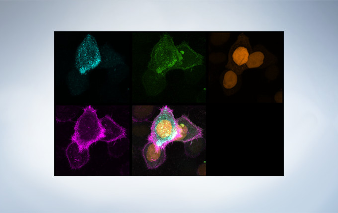
Image data courtesy of Dr. Kazuhiro Aoki, Dr. Michiyuki Matsuda, Graduate School of Medicine, Kyoto Un
See your data unfold in real time with the live 3D image display function of the FV3000 software. 3D images can be constructed during image acquisition and shown as live images.
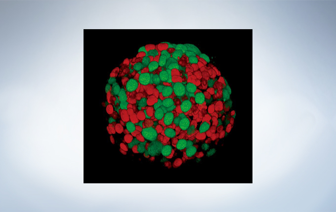
Yuji Mishima, Ph.D., Kiyohiko Hatake M.D., Ph.D. Clinical Chemotherapy, Cancer Chemotherapy Center, Japanese Foundation for Cancer Research.
Finding areas of interest in samples can be challenging. The confocal optical design of the FV3000 series supports macro to micro imaging from 1.25X up to 150X, so users can quickly switch from low magnification overview observation to high-magnification, detailed observation of regions of interest. Users can employ image stitching at both macro and micro levels to generate overview images that show samples in context.
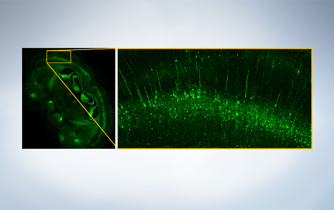
Image data courtesy of Takako Kogure and Atsushi Miyawaki, Cell Function Dynamics, Brain Science Institute of RIKEN.
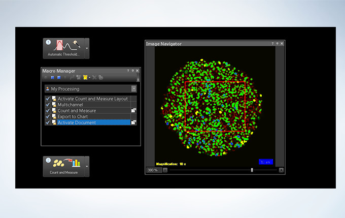
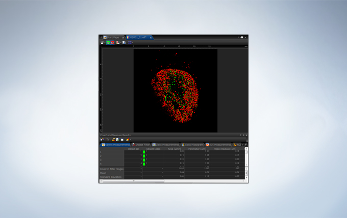
Powerful One-Click Macro Analysis with cellSens
Images alone are not enough; with integrated cellSens Count and Measure analysis, the FV3000 Series can optimize images with deconvolution and analyze them with one-click macro functionality for a broad range of morphological measurements.
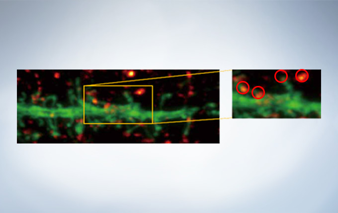
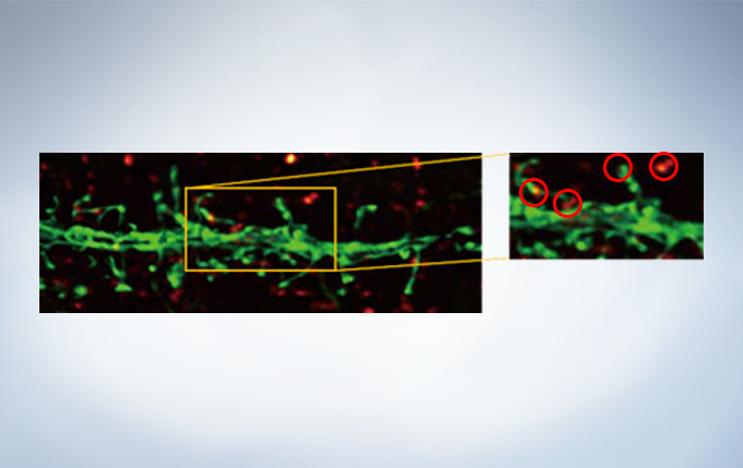
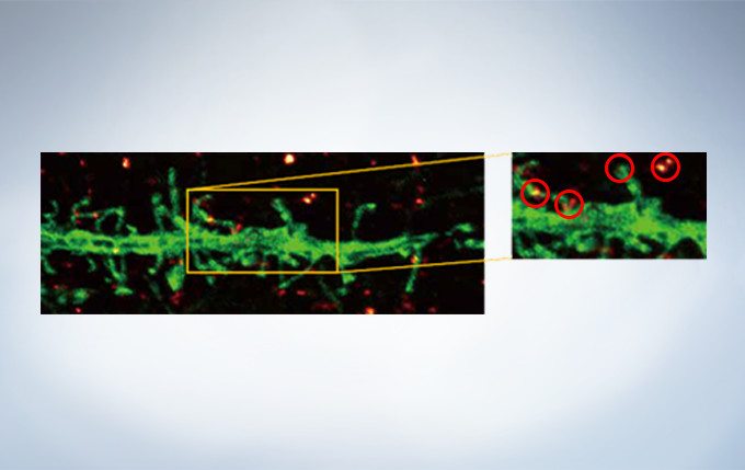
Olympus’ widely applicable super resolution method requires no special fluorophores and works for a wide range of samples. Ideal for colocalization analysis, the Olympus Super Resolution imaging module can acquire four fluorescent signals either sequentially or simultaneously with a resolution of approximately 120 nm*, nearly doubling the resolution of typical confocal microscopy. The imaging module is easy to use with minimal user training and can be added to any confocal system, making it a truly accessible method for achieving super resolution.
* Subject to objective magnification, numerical aperture, excitation and emission wavelength, and experiment conditions.
Secondary antibody labels against GFP (Alexa Fluor 488, neurons) and SV2 (Alexa Fluor 565, red). Sample courtesy of Dr. Ed Boyden and Dr. Fei Chen, MIT.
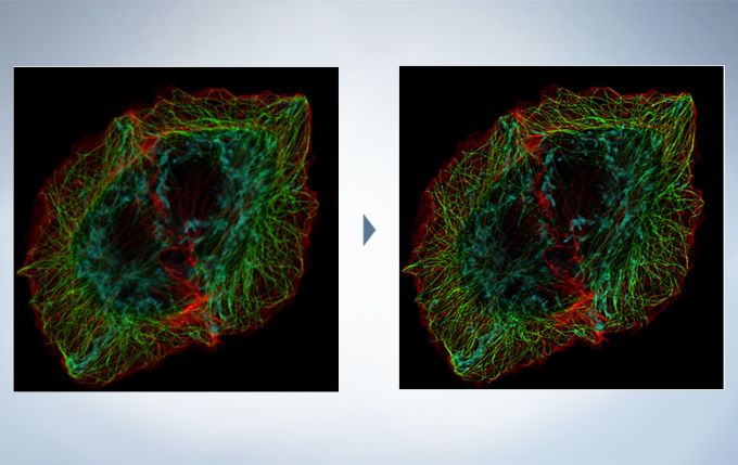
The optional constrained iterative deconvolution function improves the resolution, contrast, and dynamic ranges of confocal images obtained by the FV3000. The deconvolution function can be combined with Olympus Super Resolution (FV-OSR) to improve the z-axis resolution of the deconvolved images.
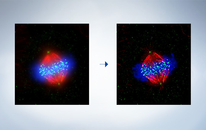
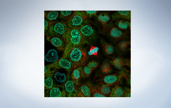
Cell line: HeLa (human cervical cancer cell line) Immunostaining: Hec1 staining (green, Alexa Fluor 488), α-tubulin staining (red, Alexa Fluor 568),DAPI staining (blue) Mitotic spindle and kinetochores are stained with anti-α-tubulin (red) and anti-Hec1 (green) antibodies, respectively. Chromosomes interact with microtubules of the mitotic spindle via kinetochores (protein structures assembled on the centromere region of chromosomes.) Image data courtesy of Masanori Ikeda and Kozo Tanaka, Department of Molecular Oncology, Institute of Development, Aging and Cancer, Tohoku University.
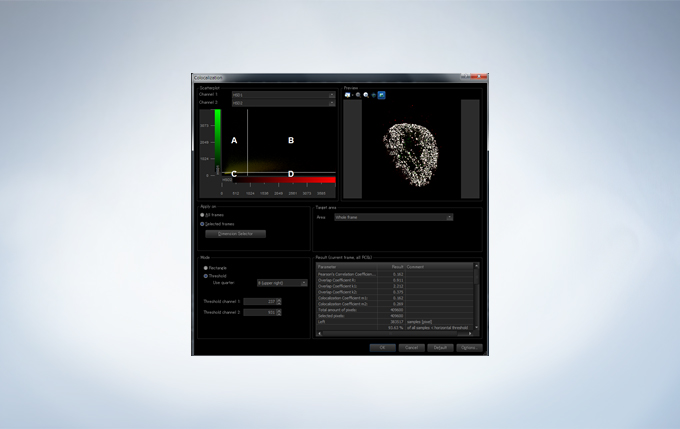
The FV3000 incorporates various optional analysis functions to complete the workflow from image acquisition through data analysis. The Count and Measure solution enables the measurement of the number, size, luminosity, and morphology of the segments. Colocalization enables the analysis of overlapping fluorescent spectra.
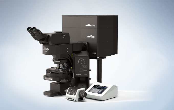
Users have their choice of two different types of scan units: galvanometer only with the FV3000 or hybrid galvanometer/resonant with the FV3000RS. The hybrid scan unit has a galvanometer scanner for high-precision scanning, as well as a resonant scanner that is ideal for high-speed imaging. With the galvanometer scanner and Olympus super resolution technology (FV-OSR), users can obtain resolutions down to 120nm with a high signal-to-noise ratio. The galvanometer scanner also features flexible scanning options, including precise tornado scanning as well as multipoint stimulation with 100ms switching time. The galvanometer scanner can image up to 16 frames per second. By switching to the resonant scanner, users can capture 30 frames per second with a full field of view at 512 x 512 pixels. By clipping down to 512 x 32 pixels, the resonant scanner can capture up to 438 frames per second to capture critical live physiological events such as calcium ion flux.
No Compromise between Speed and Field of View
Many high-speed scanning methods restrict the field of view, limiting their usefulness for examining large areas with multiple cells. The FV3000 series’ resonant scanner maintains a full 1X field of view, even at a video rate of 30 frames per second. B clipping the Y axis, additional speeds up to 438 frames per second can be achieved.
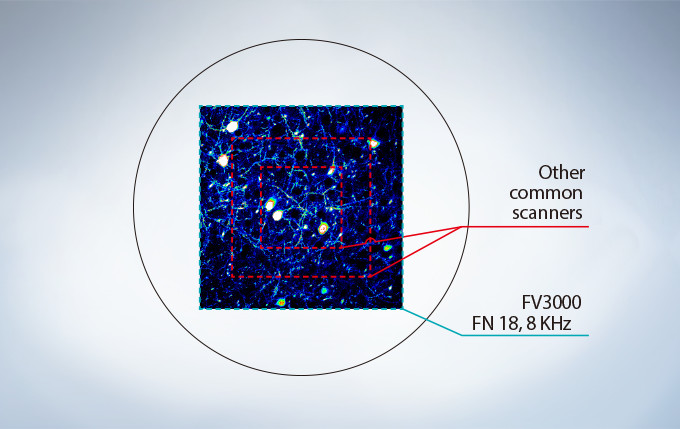
Most resonant scanners force a trade-off between speed and field of view. FLUOVIEW systems are optimized to maintain the field of view with even signal intensity so dynamic samples (e.g. calcium imaging) can be seen in the broad context of their cells and tissues.
The image above shows examples of the clipped fields of view required in other resonant scanning systems.
Platelets bound to a thrombosis in the blood vessel of a mouse. Images taken at 30 fps in full frame by resonant scanner with 2 CH GaAsP PMTs.
Image data courtesy of Dr. Takuya Hiratsuka, Dr. Michiyuki Matsuda, Graduate School of Biostudies, Kyoto University.
Optimized for Live Cell Imaging
Resonant scanning greatly reduces photobleaching and phototoxicity compared to standard galvanometer scans by preventing the excitation of fluorophores into triplet states that create reactive oxygen species. These features make live cell experiments more robust and reliable. The FV3000 series has complete laser intensity control from low to high range, enabling the system to use the minimum required amount of laser power on samples. The optional laser power monitor provides consistent laser power during long-term time-lapse imaging across multiple days.
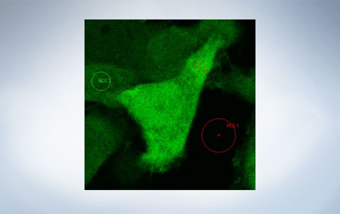
The FV3000’s ratio imaging analysis function includes an Intensity Modulated Display (IMD) function in the software that displays quantitative fluorescence ratio changes during both standard and high-speed acquisitions. This function is particularly useful for calcium and FRET imaging where a pure ratio display provides poor contrast in background areas.
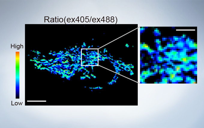
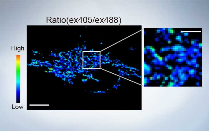
sGFP1-mito reveals heterogeneity in mitochondrial thermogenesis in HeLa cells. The images of ratio (ex 405 nm/ex 488 nm) in tsGFP1-mito-expressing cells before and after CCCP treatment at 37 °C. Scale bars indicate 10 μm (whole image) and 3 μm (inset). Image data courtesy of Shigeki Kiyonaka Ph,D, Yasuo Mori Ph,D Molecular Biology Field, Department of Synthetic Chemistry and Biological Chemistry, Kyoto University.
Cardiomyoctye Image data courtesy of Yusuke Niino and Atsushi Miyawaki, Cell Function Dynamics, Brain Science Institute of RIKEN.
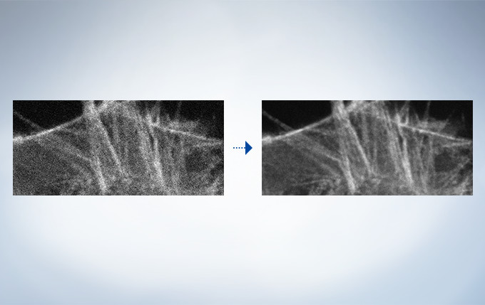
High-speed scanning at low laser power to avoid phototoxicity often decreases the signal-to-noise ratio. With rolling average post-processing, users have the flexibility to adjust high-speed time-lapse images while maintaining the time scale and keeping the original data.
In time-lapse imaging, moving objects can be automatically detected, tracked, and analyzed. cellSens software’s tracking function provides a powerful and intuitive tool to quantify dynamic processes such as cell movement and division.
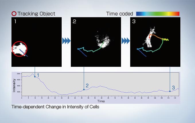
Image data courtesy of: Kazuhiro Yagita, M.D. Ph.D. Department of Physiology and Systems Bioscience, Kyoto Prefectural University of Medicine
Reference: Proc Natl Acad Sci U S A. 107(8): 3846–3851(2010)
https://static1.olympus-lifescience.com/data/Video/Library/cellSens_tracking-web2_406.mp4?rev=3464
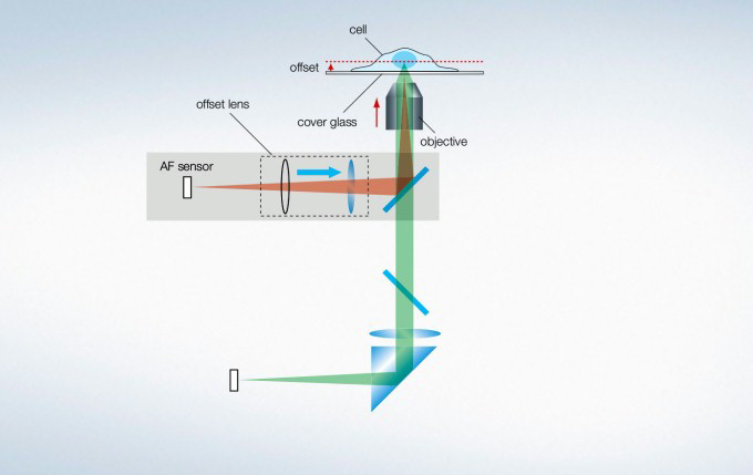
The IX3-ZDC2 Z-drift compensator uses minimally-phototoxic infrared light (laser class 1) to identify the location of the sample plane. One-shot autofocus (AF) mode enables several focus positions to be set as desired for deeper samples, enabling efficient Z-stack acquisitions in multiposition experiments. The continuous AF mode keeps the desired plane of observation precisely in focus, avoiding focus drift due to temperature changes or the addition of reagents, making it ideal for measurements that require more stringent focusing. Furthermore, the increased optical offset enables continuous AF with plastic vessels or with dry objectives. The Z-drift compensator is also compatible with silicone objectives (in AF mode).
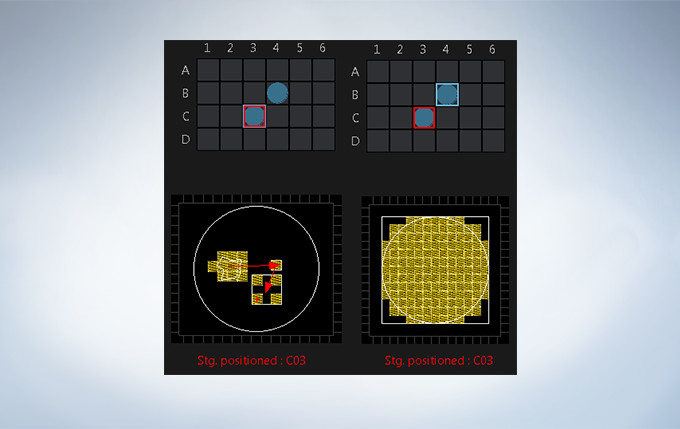
Multi-area time-lapse and stitching provide robust and accurate time-lapse data, and enable users to generate detailed overview images to see their data in context. The well navigator function provides sophisticated, intuitive controls for a wide range of cell culture vessels and custom plates.
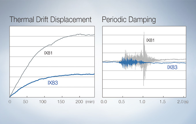
A Z-drive guide installed near the revolving nosepiece combines high thermal rigidity with the stability of a wraparound structure to significantly reduce the impact of heat and vibration and improve the quality of time-lapse imaging.
The microscope comes equipped with a hard-disk drive (HDD) recording function. The images are stored automatically in the HDD. Large volumes of data, such as those obtained from long-term time-lapse imaging, can be easily collected.
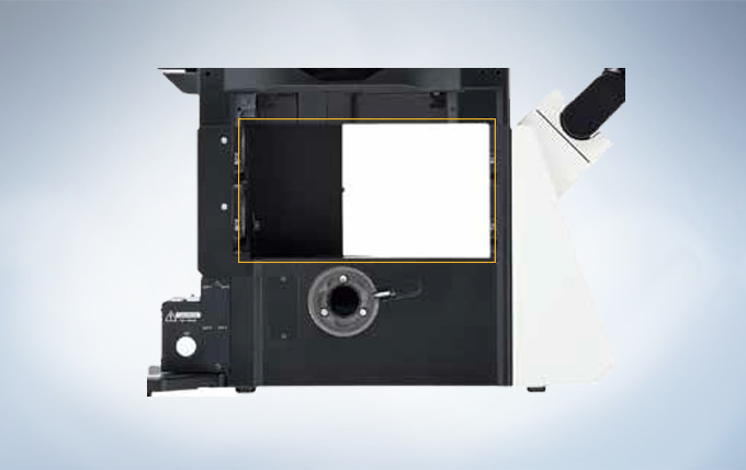
The umbra unit is designed specifically for fluorescence observation under bright room conditions. It efficiently blocks out room light, enhances the contrast of fluorescence, and enables clear fluorescence observation without the need for a dark room.
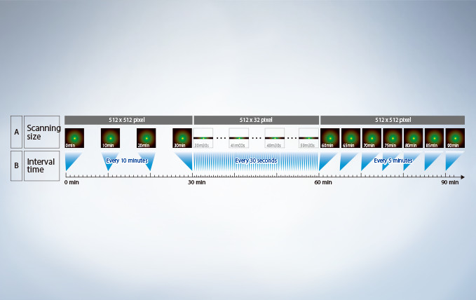
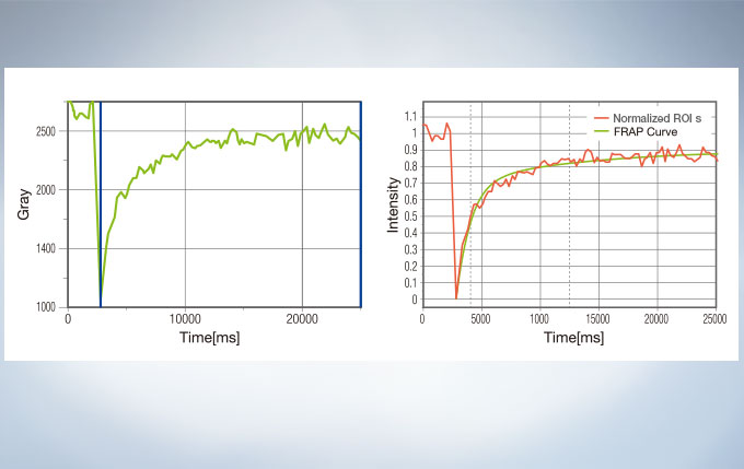
With the Sequence Manager software module, complex protocols are handled with ease and accurate timing. Multi-day time-lapse experiments are controlled with microsecond scan accuracy and millisecond sequence execution accuracy. Various protocols, such as time-lapse with different time intervals, switching between high and low magnification, and photo-stimulation between imaging by FRAP or FRET (acceptor photobleaching), can be performed.
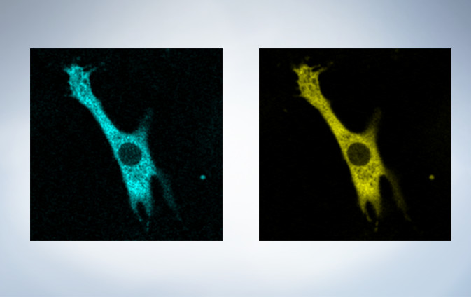
The cellSens Life Science Analysis module enables analysis of images from FRAP or FRET experiments. In FRAP, τ/2 and the Mobile/ Immobile fraction can be estimated by fitting the curve of luminosity change caused by fluorescence recovery after bleaching. FRET enables the measurement of FRET efficiency by acceptor photobleaching, ratio imaging, and sensitized emission.
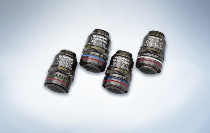
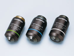
Olympus offers four high NA silicone immersion objectives that deliver excellent performance for live cell imaging. The refractive index of silicone oil (ne≈1.40) is close to that of living tissue (ne≈1.38), enabling high-resolution observations deep inside living tissue with minimal spherical aberration caused by refractive index mismatch. Silicone oil does not dry out or harden, so there is never a need to refill oil, making it ideal for extended time-lapse observations.
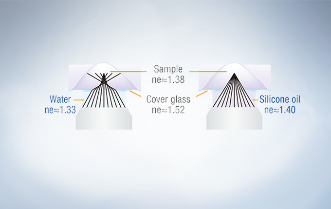
In deep tissue observation, image quality depends on keeping the refractive index of the sample and immersion medium as close to each other as possible. When working with a silicone immersion objective, the difference between the refractive index of the samples and silicone oil is minimal, thus enabling brighter fluorescence images with higher resolution for deep tissue observation.
UPLSAPO30XS: For a broader view and greater depth
Magnification: 30X, NA: 1.05 (silicone oil immersion), W.D.: 0.8 mm,
cover glass thickness: 0.13 – 0.19 mm, operating temperature: 23 – 37 °C
UPLSAPO40XS : For a good balance between field of view and resolution
Magnification: 40X, NA: 1.25 (silicone oil immersion), W.D.: 0.3 mm,
cover glass thickness: 0.13 – 0.19 mm, operating temperature: 23 – 37 °C
UPLSAPO60XS2: For 3D observations with superior resolution
Magnification: 60X, NA: 1.30 (silicone oil immersion), W.D.: 0.3 mm,
cover glass thickness: 0.15 – 0.19 mm, operating temperature: 23 – 37 °C
UPLSAPO100XS: For greater brightness at depth in closely defined regions
Magnification: 100X, NA: 1.35 (silicone oil immersion), W.D.: 0.2 mm, cover glass thickness: 0.13 – 0.19 mm, operating temperature: 23 – 37 °C
This oil immersion objective minimizes lateral and axial chromatic aberration in the 405–650 nm spectrum. Colocalization images are acquired reliably and images are measured with superior positional accuracy. The objective also compensates for chromatic aberration through near infrared up to 850 nm, making it the ideal choice for quantitative imaging.
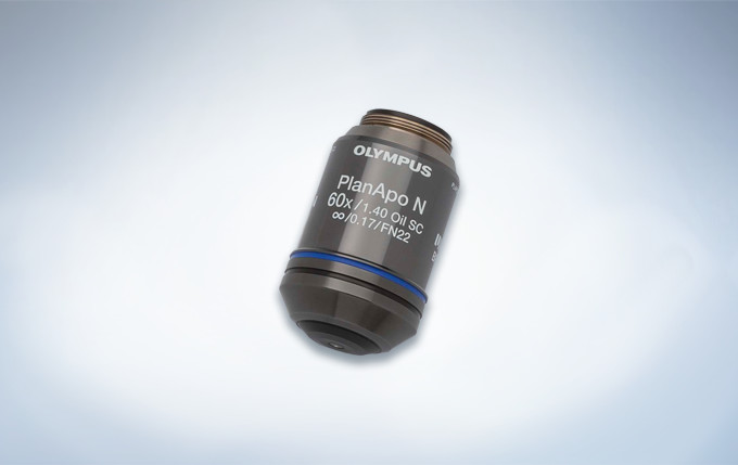
Magnification: 60X NA: 1.4 (oil immersion)
W.D.: 0.12 mm
Chromatic aberration compensation range: 405 – 650 nm
Optical data provided for each objective.
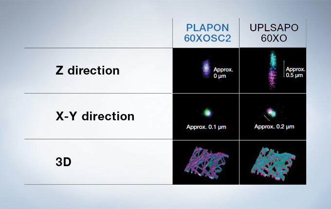
Performance Comparison of the PLAPON60XOSC2 and the UPLSAPO60XO
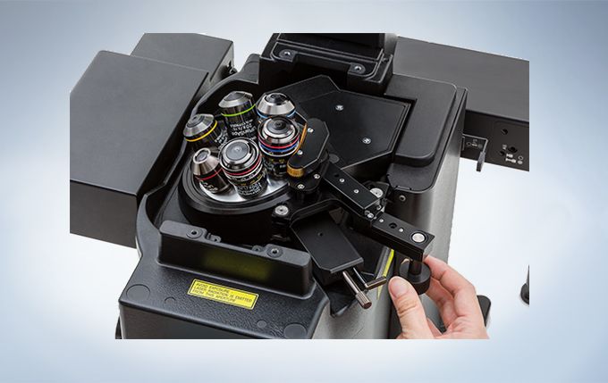
The correction collar adjusts the lens position of objectives to correct the spherical aberration caused by refractive index mismatch, resulting in the improvement of image quality, such as resolution, brightness and contrast. The correction collar is especially necessary for objectives with high NA when they are used for super resolution imaging, because they are greatly affected by spherical aberration. The remote correction collar unit is useful for easy adjustment and improvement of the image quality, and operable on all UIS2 objectives which have a correction collar.
Customizable and saveable layouts make it easy to tailor the interface to your workflow and experiment needs, from basic to complex.
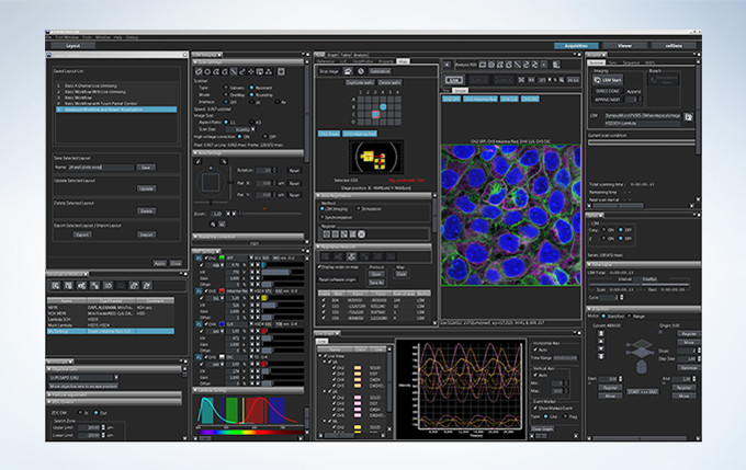
Layout
Start by selecting your preferred display with specific tools for basic to complex acquisition.
Acquisition Condition
Reload settings that were ideal for your last experiment to provide consistency.
Acquisition
Activate basic to complex acquisitions with live ratio, intensity modulated display, quantitative region of interest (ROI) graphing or spectral unmixing display, and data backup for added security.
Viewer
Review data as it is generated. Generate 3D and 4D views and animations to explore and share data in depth.
Analysis
Extract data from images using online or offline processing. Analytical tools include Olympus super resolution technology (FV-OSR) and powerful cellSens software with features such as deconvolution, filtering, count and measure, and one-click macros.
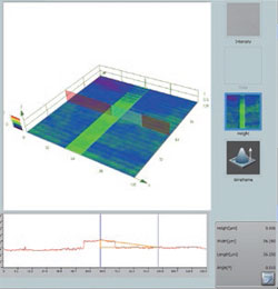
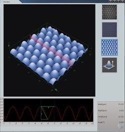
LEXT OLS4500은 광학 현미경, 레이저 현미경(LSM), 프로브 현미경(SPM)을 한 대에 결합한 복합 현미경입니다. 관찰 배율은 수십 배에서 수백만 배까지 광범위한 영역을 커버하고 관찰 포인트를 잃는 일 없이 원활하게 밀리미터부터 나노미터까지 관찰 및 측정이 가능합니다.
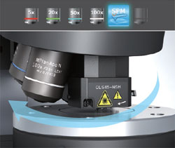
저배율에서 고배율 관찰이 가능한 4 개의 대물 렌즈와 SPM 장치를 전동 리볼버에 장착하여 배율과 관찰 방법을 원활하게 전환 할 수 있어 관찰 대상을 잃을 일이 없이 바로 나노미터까지 검색 할 수 있는 현미경입니다.
첨단 광학 기술이 뒷받침된 다양한 관찰 방법과 저배율부터 고배율까지 관찰 대상을 쉽게 찾을 수 있습니다. 또한 광학 현미경으로는 보이지 않는 관찰 대상을 레이저 현미경으로 찾을 수 있습니다. 레이저 미분 간섭 (DIC) 관찰에서는 나노 수준의 미세 요철의 라이브 관찰도 가능합니다.
샘플 세팅 이후의 모든 작업을 1대의 장치로 수행 할 수 있습니다.
관찰대상을 신속, 정확하게 SPM 현미경 모드로 가져올 수 있기 때문에, 한 번의 스캐닝 영역 내에서 원하는 SPM 이미지를 얻을 수 있습니다.
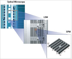 OLS4500은 광학 현미경, 레이저 현미경, 프로브 현미경의 일체형이기 때문에 샘플을 옮길 필요 없이 세 가지의 현미경을 자유 자재로 전환하며 관찰 및 평가가 가능합니다. 각각이 가진 뛰어난 기능으로 효율적인 최적의 결과 값을 얻을 수 있습니다.
광원으로 백색 LED를 사용하기 때문에, 색 재현성이 뛰어난 고해상도 컬러 이미지를 볼 수 있습니다. 4개의 대물 렌즈로 저배율에서 고배율까지 관찰이 가능합니다. OLS4500은 광학 현미경의 기능을 최대한 활용하여 가장 많이 사용되는 명시야관찰 (BF)을 비롯해 미세한 요철에 컨트라스트를 넣어 입체적으로 시각화하는 미분간섭관찰 (DIC), 샘플의 편광특성이 색으로 표현되는 간이 편광 관찰이 가능합니다. 또한 노출시간을 바꾸어 여러장의 이미지를 촬영, 합성 하는 것으로 밸런스가 좋은 밝기와 향상된 텍스처의 이미지를 보여주는 HDR기능(High Dynamic Range)을 이용 할 수 있습니다. 다양한 관찰 방법으로 신속하게 관심 영역을 찾을 수 있습니다.
가장 널리 사용되는 관찰 방법입니다.
자연스러운 이미지와 사실적인 칼라 재현. 컨트라스트가 있는 샘플의 관찰에 적합합니다.
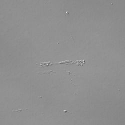
Differential Interference Contrast
명시야에서는 보이지 않는 샘플의 미세한 단차를 시각화합니다. 금속조직, 하드디스크 및 웨이퍼 연마 표면 같은 거울위의 스크레치나 이물질 관찰에 적합합니다.

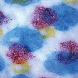
편광 (특정 진동 방향을 갖는 빛)을 조사하여 샘플의 편광 특성 (굴절률 등)을 시각화합니다. 금속 조직, 무기물, 반도체 재료 등의 관찰에 적합합니다.
노출 시간을 바꾸어 여러 장의 사진을 찍어 합성하는 것으로, 밝은 부분, 어두운 부분을 균형있게 볼 수 있습니다. 또한 텍스처 (표면 상태)를 강조함으로써 세밀한 관찰이 가능합니다.
405nm 짧은 파장의 레이저 광원과 높은 N.A의 전용 대물렌즈를 사용하여, 높은 평면 분해능을 가집니다. 광학 현미경으로는 보이지 않았던 관찰대상을 선명한 이미지로 관찰하는 것이 가능합니다. 레이저 미분간섭(DIC)은 나노미터의 마이크로영역 표면의 실시간 관찰도 가능합니다.
저배율에서 고배율 관찰이 가능한 4개의 대물 렌즈와 소형 SPM 장치를 전동 리볼버에 장착. 광학현미경 또는 레이저 현미경 50배, 100배의 실시간 관찰에서는 SPM 스캔범위가 시야의 중심에 표시되므로 관찰 포인트를 이 위치에 맞춘 후, 프로브현미경으로 전환 하는 것만으로 관찰대상에 정확하게 접근 할 수 있습니다. 따라서 원하는 이미지를 한번의 SPM 스캔으로 획득할 수 있고, 작업의 효율성과 캔틸레버의 소모를 줄일 수 있습니다.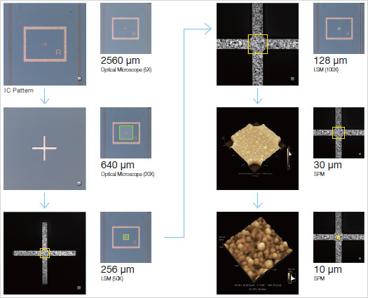
 캔틸레버 설치, 스캔 범위 설정 등 프로브 현미경으로 관찰하는 데 필요한 준비는 안내 화면에 따라 수행 할 수 있어, 경험이 적은 사람이라도 안심하고 작업을 할 수 있습니다.
OLS4500에서는 전동 리볼버에 장착하는 대물 렌즈 형 SPM 헤드를 사용. 대물 렌즈와 캔틸레버 끝을 동축, 동초점으로 배치하여, SPM 관찰로 전환 하더라도 관찰 포인트를 잃지 않습니다. 또한 새로 개발한 소형 SPM 헤드는 강성을 높여 기존제품보다 이미지의 노이즈를 줄이고 응답성을 향상 시켰습니다.
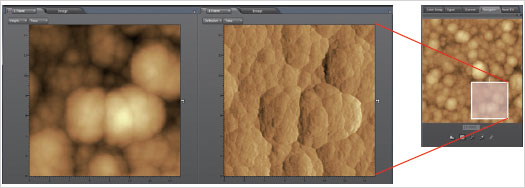
네비게이터 기능은 프로브 현미경에서 얻은 이미지의 필요한 부분을 더 크게 확대하여 관찰 할 수 있습니다. 이미지에서 확대 범위를 커서로 설정하여 스캔을 시작하는 것 만으로 원하는 SPM 이미지를 얻을 수 있습니다. 스캔 범위는 자유롭게 설정할 수 있으므로 관찰 · 측정 효율성이 크게 향상됩니다.
각종 측정 모드에서 얻은 이미지는 목적에 따른 분석이 가능하며, 측정 결과는 CSV 형식으로 출력 할 수 있습니다. OLS4500는 다음의 분석 기능이 있습니다.
캔틸레버와 샘플 사이에서 작동하는 반발력이 일정하게 되도록 제어하면서 캔틸레버를 정적으로 스캔하며 샘플의 높이를 이미지화 시키다. 힘 곡선 측정에도 사용 할 수 있습니다.
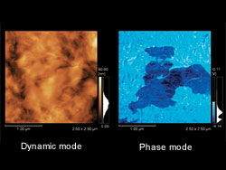
캔틸레버를 공진 주파수 근처에서 진동시켜 진폭이 일정하게 되도록 Z축 방향의 거리를 제어하는 것으로, 샘플의 높이를 이미지화 시킨다. 특히 고분자 화합물 같은 부드러운 표면 샘플 및 점착성이 있는 샘플에 적합합니다.
다이나믹 모드에서 스캐닝하는 동안 캔틸레버 진동의 위상 지연을 감지합니다. 샘플 표면의 물리적 특성의 차이를 이미지화 할 수 있습니다.

샘플에 바이어스 전압을 인가하여 캔틸레버와 샘플 사이에 흐르는 전류를 감지하여 이미지화시킵니다. 또한 I / V 측정도 가능합니다.
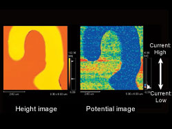
전도성 캔틸레버를 이용하여 교류 전압을 인가하여 캔틸레버와 샘플 표면 사이에 작용하는 정전기의 힘을 감지하고 샘플 표면의 전위를 이미지화 시킵니다. KFM (Kelvin Force Microscope)라고도합니다.
자력을 갖는 캔틸레버를 위상 모드에서 검사하여, 진동 하는 캔틸레버의 위상 지연을 감지하고 샘플 표면의 자기 정보를 이미지화 시킵니다. MFM (Magnetic Force Microscope)라고도 합니다.
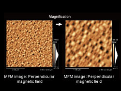
OLS4500의 높은 해상도와 405nm의 광학시스템에 맞게 설계된 전용렌즈의 채택으로 기존에 측정 불가능했던 급경사면의 이미지를 손쉽게 취득할 수 있습니다.
405nm의 단파장 레이저 빛과 높은 N.A의 전용 대물 렌즈 사용으로 최대 0.12μm의 평면 분해능을 실현. 샘플 표면의 서브 마이크론 측정이 가능합니다. 또한 고정밀 리니어 스케일과 올림푸스 만의 밝기 감지 기술은 서브 마이크론에서 수백 마이크론의 높이 차이를 감지 할 수 있습니다. 또한 레이저 현미경에 의한 측정은 측정기 2 가지 지표인 ‘정확도'(참값에 근접)과 ‘반복성'(편차의 작음) 모두의 성능을 보장하고 있습니다.

고배율의 이미지에서는 시야범위가 좁아지지만, 스티칭기능으로 최대625장까지 이미지를 붙여 높은 분해능과 넓은 시야범위로 이미지 데이터를 얻을 수 있습니다. 또한 넓은 시야 이미지로 3D 디스플레이 및 3D 측정이 가능합니다.

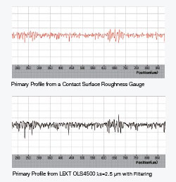 최근 산업 제품의 크기와 무게의 지속적인 감소로, 이를 구성하는 부품도 소형화 되고 있습니다. 이러한 경향은 표면 거칠기 측정뿐만 아니라, 형상 측정에서도 중요성이 증가하고 있습니다. 이러한 시장의 요구를 반영하여 ISO에서 규정하는 3D 표면 질감 측정 장치 (ISO 25178-6)의 목록에 LSM과 AFM을 추가했습니다. 이 비접촉 표면 거칠기 측정은 (삭제) 기존의 접촉 표면 거칠기 측정기와 같은 공식적인 평가 기준으로 인정된다는 것을 의미합니다. OLS4500은 ISO에 적합한 거칠기 파라미터를 제공합니다.
비접촉 표면 거칠기 측정은 평면 거칠기뿐만 아니라 선 거칠기를 얻을 수 있습니다.
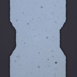
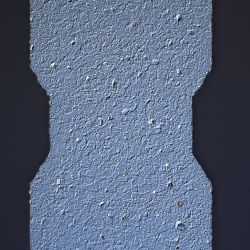
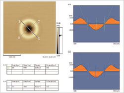
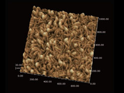
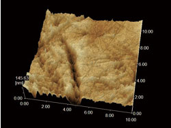
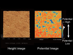
표면 거칠기 측정은 샘플 표면에서 설정 한 영역의 분포와 특징을 파악할 수 있으며, 3D 이미지와 대조 한 평가가 가능합니다. OLS4500은 LSM 또는 SPM 기능을 사용하여 표면 거칠기를 측정 할 수 있습니다. 이 두 기능은 샘플 속성 혹은 관찰 목적에 따라 구분하여 사용할 수 있습니다.
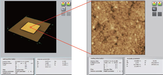
(우) 프로브 현미경에 의한 표면 거칠기 (10μm x 10μm)
OLS4100은 접촉식 표면 거칠기 측정기와 같은 거칠기 (2 차원) 파라미터를 보유하고 있습니다. 접촉식 표면 거칠기 측정기와 같은 조작성, 호환 측정 결과를 얻을 수 있습니다.
OLS4500은 ISO25178 규격 거칠기 (3 차원) 파라미터를 보유하고 있습니다. 평면 영역에서 평가를 실시하는 것으로, 높은 신뢰성이 있는 분석이 가능합니다.
가시 광선 영역 (400-800 nm의 파장)을 사용하여 광학 현미경 이미지를 1000배 정도의 배율로 관찰 할 수 있습니다. 광학 현미경의 특징은 샘플이 가지는 색을 그대로 관찰 할 수 있으며 관찰 방법을 바꾸는 것으로 요철을 강조하거나 물질의 특성 (편광 특성)을 이용한 관찰을 할 수 있다는 것 입니다. OLS4500에서는 다음 관찰 방법이 가능합니다.
광학 현미경의 평면 분해능은 사용하는 빛의 파장에 크게 의존합니다. 단파장의 레이저 빛을 사용하는 레이저 현미경은 가시 광선을 사용하는 기존의 현미경에 비해 평면 분해능이 뛰어납니다. OLS4500는 405nm의 단파장 반도체 레이저를 사용하며, 높은 개구 수 (NA) 전용 대물 렌즈, 공 초점 광학계를 결합하여 최대 0.12μm의 평면 분해능을 실현하고 있습니다. 또한 올림푸스 만의 2 차원 스캐너에 의한 XY 스캐닝 기능에서 최대 4096 픽셀 x 4096 픽셀의 고해상도 스캔을 가능하게 하고 있습니다.
레이저 현미경은 단파장 반도체 레이저와 공 초점 광학계를 사용하여 초점이 맞는 반사광을 감지하고 초점이 맞지 않는 부분의 반사광은 제외됩니다. 정밀 리니어 스케일과 결합하여 정확한 3 차원 측정이 가능합니다.
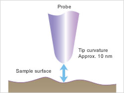
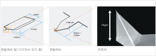
끝 곡률이 10nm 정도의 미세한 탐침 (프로브)을 샘플 표면에 접근 시켜 샘플 사이에 발생하는 역학적 · 전기적 상호 작용을 감지하면서 스캐닝하여 3 차원 적으로 관찰하는 현미경을 총칭하여 프로브 현미경 (SPM)라고 합니다. 대표적인 것으로 탐침과 샘플 표면 사이에 작용하는 인력과 척력을 감지하여 스캔 이미지를 얻는 원자 힘 현미경 (AFM : Atomic Force Microscope)이 있습니다. 나노 수준에서 관찰하는 것으로, 샘플의 모습을 세밀하게 파악할 수 있습니다.
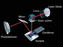
OLS4500은 끝에 탐침 (프로브)을 배치 한 캔틸레버의 미세한 굴곡 량 (변위)를 고감도로 검출하는 광 지렛대 방식을 사용. 레이저 빛을 캔틸레버 후면에 반사시켜 광 검출기의 일정한 위치에 맞게 압전 소자에서 Z축으로 구동시켜 미세한 Z축 방향의 변위를 정확하게 읽습니다.
프로브 현미경의 다양한 모드는 샘플 표면 형상 관찰, 측정, 또한 물리적 특성의 분석이 가능합니다. OLS4500는 다음 모드를 지원합니다.
탐침 (프로브)은 길이 100μm에서 200μm 정도의 얇은 판 모양 캔틸레버 끝으로 형성되어 있습니다. 캔틸레버는 샘플에 따라 용수철 상수, 공진 주파수를 선택합니다. 스캔 반복에 따라 탐침 (프로브)은 마모하기 때문에 필요에 따라 혹은 정기적으로 캔틸레버 팁을 교체합니다.