카테고리 보관물: Microscope

Ease of Use

Comfortable and Easy to Use


Traditional Techniques Made Easy: Simple Illuminator

The illuminator minimizes complicated actions that are usually necessary during microscope operation. A dial at the front of the illuminator enables the user to easily change the observation method. An operator can quickly switch between the most frequently used observation methods in reflected light microscopy, such as from brightfield, to darkfield, to polarized light, in order to readily change between different types of analysis. In addition, simple polarized light observation is adjustable by rotating the analyzer.

An operator can quickly switch between frequently used observation methods.(Polished sample of AlSi)

Intuitive Microscope Controls: Simple FS and AS Settings

Using the proper aperture stop and field stop settings provides good image contrast and makes full use of the numerical aperture of the objective. The legend guides the user to the correct setting based on the observation method and objective in use.


Find the Focus Quickly: Focus Scale Index

The focus scale index on the frame supports quick access to the focal point. Operators can roughly adjust the focal point without viewing the sample through an eyepiece, saving time when inspecting samples that are different heights.


For Consistent Illumination: Light Intensity Manager

During the initial setup, the illumination intensity can be adjusted to match the specific hardware configuration of the coded illuminator and/or coded nosepiece.

Conventional Light Intensity

Similar to conventional microscopes, the image appears darker with increasing magnification and observation methods that consume more light

Light Intensity Manager

The microscope automatically adjusts the light intensity to the correct value when changing magnification or observation method


Easy and Ergonomic Operation

Ergonomics are of the utmost importance for all users. Both standalone microscope users and those integrating with OLYMPUS Stream image analysis software benefit from ergonomic handset controls that clearly display the hardware position. The simple handsets enable the user to focus on their sample and the inspection they need to perform.


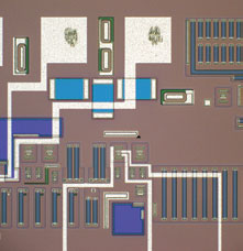
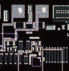
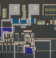
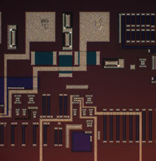
A System Designed for Image Analysis

For Restoring Microscope Settings: Coded Hardware

The BX3M employs new coded functions that integrate the microscope’s hardware settings with OLYMPUS Stream image analysis software. The observation method, illumination intensity, and objective position are all recorded within the software and/or the handset. The coded functions enable the microscope settings to be automatically saved with each image, making it easier to reproduce the settings at a later time and provide documentation for reporting purposes. This saves the operator time and minimizes the chance that an incorrect setting will be used. The current observation settings are always clearly displayed both on the hand switch and in the software.

BX53M

BX53M

Ease of use Feature Image quality Configuration

Designed with modularity in mind, the BX3M series provide versatility for a wide variety of materials science and industrial applications. With improved integration with OLYMPUS Stream software, the BX3M provides a seamless workflow for standard microscopy and digital imaging users from observation to report creation.


User-Friendly

  • Traditional Techniques Made Easy: Simple Illuminator
  • Intuitive Microscope Controls: Simple FS and AS Settings
  • Find the Focus Quickly: Focus Scale Index
  • Consistent Illumination: Light Intensity Manager
  • Easy and Ergonomic Operation
  • Easy Restore Microscope Settings: Coded Hardware
  • Basic Measurement Functions

Functional

  • The Invisible Becomes Visible: MIX Observation
  • Create All-in-focus Images: EFI
  • Easily Move the Stage for Panorama: Instant MIA
  • Capture Both Bright and Dark Areas: HDR
  • Adaptable to Suit Observational and Analysis Preferences
  • Accommodates a Wide Range of Samples

Precision Optics

  • Superior Optical Performance: Wave Front Aberration Control
  • Stable Color Temperature and High-Intensity White LED Illumination
  • Support Precise Measurement: Auto Calibration
  • Seamless Stitching : Image Shading Correction

Fully Customizable

  • Various configurations to meet users’ requirements
  • Modular Design, Build Your System Your Way

SZX10

SZX10

OLYMPUS SZX10은 Working Distance와 Field Size가 중요할 때 선택하시는 모델입니다. 렌즈의 디자인에 따라 왜곡현상없이 제품 혹은 샘플의 고유 색감을 그대로 관찰하실 수 있습니다.

  • 안정성 및 반복성
  • 인체공학적이고 빠른 사용
  • 지능적인 디지털 영상

안정성 및 반복성

 빠른 검사가 가능한 조절 손잡이

Zoom knob with click-stop

0.63배 에서 6.3배에 이르는 넓은 범위의 배율은 듀얼 렌즈 홀더를 통해 더 높은 배율도 볼 수 있으며, 별도의 렌즈 교체 없이 다양한 배율에서의 관찰이 가능합니다. 축을 이루는 빛 경로 설정의 장점은 카메라로 찍은 측정값이 현미경에서 샘플의 방향에 상관없이 모든 방향으로 안정적이고 정확하며 많은 영상 및 측정 작업들은 같은 배율에서 일관성있고 비교할 수 있는 정확한 결과값을 보장합니다. 11개의 click-stop 지점들은 중요한 기능들을 빠르고 쉽게 사용할 수 있도록 합니다.


8개의 호환가능한 렌즈로부터의 뛰어난 성능

Lineup of Objective Lenses

DFPL2x/DPFL1.5x /DFPL 0.75x /DFPL 0.5x : DFPL 렌즈 시리즈는 뛰어난 색 재현성과 샘플의 모양을 정확하게 구현합니다.

DFPLAPO 1.25x /DFPLAP01x4 : 색 수차 및 구면 수차를 제거한 가장 높은 성능의 렌즈로 뛰어난 분해능과 contrast, 이미지 평면도와 최소한의 왜곡 등의 특징을 가집니다.

SZX-ACH1.25x /SZX-ACH1x : Long Working Distance와 고분해능을 갖춘 균형잡힌 렌즈입니다.


인체공학적이고 빠른 사용


인체공학적으로 편안한 사용 환경

OLYMPUS는 인체공학적인 디자인이 사용자들에게 가장 중요한 요소라고 믿습니다. 따라서, 모든 동작은 사용하기 쉽고 눈이 편안한 접안렌즈, 낮은 위치의 스탠드 등으로 사용하는데 발생할 수 있는 스트레스를 최대한으로 줄였습니다.
SZX2 실체현미경의 고급화된 OLYMPUS Comfort View 접안렌즈는 3D 이미지를 볼 수 있도록 넓은 범위를 한 번에 볼 수 있게하며 장시간 사용시에도 편안함을 가져다 줍니다.
또한 눈의 피로를 줄이며 실체 이미지를 더 쉽게 볼 수 있게 합니다. OLYMPUS는 튜브(헤드) 부분의 각도가 5~45로 조절할 수 있어 다양한 사용자들이 만족하며 사용할 수 있습니다.


자동기능

SZX10 전동 초점 장치는 화장 초점 영상(EFI)와 완벽하게 자동화된 디지털 문서를 만들 수 있습니다. 이러한 기능은 3D 이미지를 만들 수 있으며, 문서상의 이미지와 실제 눈으로 본 이미지의 격차를 줄여줍니다. 자동 검사를 용이하게하기 위해 OLYMPUS STREAM 이미징 소프트웨어 제품군은 간단한 2D 측정부터 복잡한 위상 분석에까지 다양한 환경에 사용하기 쉬운 모든 도구들을 제공합니다.


다양한 종류의 스탠드와 조명 장치

Low-profile LED Illumination Base

OLYMPUS SZX2는 OLYMPUS LED 기반의 특별한 시스템을 갖추고 있어 투과 조명의 미세한 조정으로 밝기와 영상을 컨트롤 하실 수 있습니다. 울트라 슬림의 40mm Base와 함께 SZX2는 가장 인체공학적인 높이의 스테이지를 지원합니다. 게다가 명시야, 암시야,  Oblique 조명의 선택을 회전식 스위치를 통해 쉽게 할 수 있습니다. 긴 수명의 LED는 샘플이나 스테이지에 열을 전달하지 않으며 Material 실체현미경으로써의 유연성을 강화하였습니다.


다양한 종류의 반사형 조명 장치

링 라이트 방식의 조명이나 동축 조명등의 다양한 반사형 조명유닛들의 사용이 가능합니다. 사용자의 어플리케이션에 맞는 최적의 조명 시스템을 선택하실 수 있습니다.


지능적인 디지털 영상

디지털 영상

SZX16과 함께 당신은 샘플의 실시간 디지털 영상을 보여주거나 문서를 제작하고 싶어질 것입니다. OLYMPUS 쿨링 방식의 DP를 같이 사용하여 매우 유용한 이미징 스테이션을 구착할 수 있습니다. 이 것은 빠른 속도의 라이비 영상 관찰 뿐만 아니라 고해상도(1730)만화소까지)의 문서 제작도 가능합니다. 발전된 인터페이스는 샘플이 움직이는 동안에도 색재현성이 뛰어나고 색변화가 자유롭습니다.

OLYMPUS의 정교한 현미경 디지털 카메라 및 이미지 분석 소프트웨어 패키지는 관찰, 리포트 생성, 데이터베이스, 보관 및 SZX16 전동 줌 초점 제어를 포함한 다수를 작업을 지원합니다. OLYMPUS가 디지털 카메라 및 분석 / 측정 소프트웨어의 전체 라인을 제공하며, 고객님의 OLYMPUS 담당자는 사용자의 요구 사항에 맞춰 충족시켜드릴 수 있도록 도와 드릴 것입니다.


Work Comfortably and Productively

Natural Posture, Reduced Stress, and Increased Productivity

The ergonomic long tilting trinocular provides an optimized work position by bringing the microscope closer to the user while the extendable eyepoint adjuster provides flexibility for users of different heights. The SZX series’ ergonomic instruments reduce stress during observation by providing the most comfortable position for each user, increasing work efficiency.


Convergence Angle in Tube Relieves Eyestrain

Olympus research established a correlation between stereo microscope optical systems and eyestrain. Certain convergence angles between the left and right optical paths can cause discomfort. The SZX2 series is designed with a convergence angle that completely compensates for each optical path. This solution effectively eliminates eyestrain, even during prolonged observation.


Ergonomic Zoom and Focus Knobs for Fatigue-free Use

The position of the zoom knob, size and position of the coarse/fine focusing knob, and the fine focus stroke are designed for easy operation. The enhanced fine focus stroke results in easy and precise focusing.


Slim Design Illumination Stand for Easy Access to Samples

Illumination stands are designed to be easy to use and fatigue-free. The slim LED transmitted light illumination stand, at approximately 40 mm in height, features easily adjustable fingertip illumination control and provides easy access to samples.


Specification

Research Stereomicroscope System SZX10 Specifications
Optical System Galilean Optical System
Total Magnification 3.15x-378x*1
Zoom Body Zoom Ratio 10 (0.63x-6.3x)
AS Built-in
Observation Tube Binocular/Trinocular/Tilting Trinocular Observation Tube
Extendable Eyepoint Adjuster SZX2-EEPA
Focus Focusing Unit/Coarse Fine Focusing Unit/Heavy-duty Coarse Fine Focusing Unit/Motorized Focusing Unit
Objective Lens Magnification Type N.A. W.D. (mm) Total Magnification *2 *3 Field Diameter of View (mm)*2 *3
0.5x Plan Achromat 0.05 171 3.2x-31.5x Ø69.8-Ø7
0.75x Plan Achromat 0.075 116 4.7x-47.3x Ø46.6-Ø4.7
1x Plan Apochromat 0.1 81 6.3x-63x Ø34.9-Ø3.5
Achromat 0.1 90 6.3x-63x Ø34.9-Ø3.5
1.25x Plan Apochromat 0.125 60 7.9x-78.9x Ø27.9-Ø2.8
Achromat 0.125 68 7.9x-78.9x Ø27.9-Ø2.8
1.5x Plan Achromat 0.15 45.5 9.5x-94.5x Ø23.3-Ø2.3
2x Plan Achromat 0.2 33.5 12.6x-126x Ø17.5-Ø1.7
Dimensions (W x D x H) 285 mm x 335 mm x 400 mm
Weight 7 kg (in Standard Configularion)
Remark *1 total magnification range possible by combining an objective lens and eyepiece
*2 in the case of using eyepiece 10x
*3 SZX2-LTTR: intermediate magnification is 1.25X, SZX2-ILLC16/10: intermediate magnification is 1.5X

SZX16

SZX16


OLYMPUS SZX16은 900 line pair/mm 의 높은 분해능을 가지고 있어 고사양의 성능을 필요로 하는 어플리케이션에 사용될 수 있도록 제작되었습니다. 0.7x – 11.5x 에 이르는 줌 배율과 듀얼터렛을 사용하여 2개의 대물렌즈를 사용하실 수 있습니다.

  • 광학의 우수성
  • 인체공학적이고 빠른 사용
  • 지능적인 디지털 영상

광학의 우수성

넓은 줌 범위

0.7배에서 11.5배에 이르는 넓은 줌 범위를 갖고 있어 추가적인 대물렌즈의 변경없이 넓은 시야에서 부터 자세한 부분까지 관찰이 가능합니다.

 다양한 종류의 대물렌즈

Lineup of Objective Lenses

SDF 시리즈의 대물렌즈와 함께 비점수차를 제거하여 높은 분해능과 contrast를 가진 이미지를 관찰 할 수 있습니다.
샘플의 요구사항에 맞게 6가지 종류의 렌즈를 통해 가장 적합한 대물렌즈를 선택하여 사용할 수 있습니다.
parfocal 렌즈의 가장 넓은 범위 (0.5x, 1.0x, 1.6x, 2.0x )와 함께 큰 줌 비율은 SZX16가 macro-view부터 micro-view까지 관찰할 수 있게 합니다. 놀라운 분해능과 배율의 선택은 작업을 더 효율적으로 하고 샘플로부터 더 정확한 정보를 얻을 수 있습니다.


인체공학적이고 빠른 사용

자동기능

SZX16 전동 초점 장치는 화장 초점 영상(EFI)와 완벽하게 자동화된 디지털 문서를 만들 수 있습니다. 이러한 기능은 3D 이미지를 만들 수 있으며, 문서상의 이미지와 실제 눈으로 본 이미지의 격차를 줄여줍니다. 자동 검사를 용이하게하기 위해 OLYMPUS STREAM 이미징 소프트웨어 제품군은 간단한 2D 측정부터 복잡한 위상 분석에까지 다양한 환경에 사용하기 쉬운 모든 도구들을 제공합니다.

인체공학

OLYMPUS는 인체공학적인 디자인이 사용자들에게 가장 중요한 요소라고 믿습니다. 따라서, 모든 동작은 사용하기 쉽고 눈이 편안한 접안렌즈, 낮은 위치의 스탠드 등으로 사용하는데 발생할 수 있는 스트레스를 최대한으로 줄였습니다.
SZX2 실체현미경의 고급화된 OLYMPUS Comfort View 접안렌즈는 3D 이미지를 볼 수 있도록 넓은 범위를 한 번에 볼 수 있게하며 장시간 사용시에도 편안함을 가져다 줍니다.
또한 눈의 피로를 줄이며 실체 이미지를 더 쉽게 볼 수 있게 합니다. OLYMPUS는 튜브(헤드) 부분의 각도가 5~45로 조절할 수 있어 다양한 사용자들이 만족하며 사용할 수 있습니다.

Viewing tube (head) of adjustable angle
Adjustable observation tube(head)

다양한 종류의 스탠드와 조명 장치

OLYMPUS SZX2는 OLYMPUS LED 기반의 특별한 시스템을 갖추고 있어 투과 조명의 미세한 조정으로 밝기와 영상을 컨트롤 하실 수 있습니다.울트라 슬림의 40mm Base와 함께 SZX2는 가장 인체공학적인 높이의 스테이지를 지원합니다. 게다가 명시야,암시야, Oblique 조명의 선택을 회전식 스위치를 통해 쉽게 할 수 있습니다. 긴 수명의 LED는 샘플이나 스테이지에 열을 전달하지 않으며 Material 실체현미경으로써의유연성을 강화하였습니다.

Low-profile LED Illumination Base

다양한 종류의 반사형 조명 장치

링 라이트 방식의 조명이나 동축 조명등의 다양한 반사형 조명유닛들의 사용이 가능합니다. 사용자의 어플리케이션에 맞는 최적의 조명 시스템을 선택하실 수 있습니다.


Work Comfortably and Productively

Natural Posture, Reduced Stress, and Increased Productivity

The ergonomic long tilting trinocular provides an optimized work position by bringing the microscope closer to the user while the extendable eyepoint adjuster provides flexibility for users of different heights. The SZX series’ ergonomic instruments reduce stress during observation by providing the most comfortable position for each user, increasing work efficiency.


Convergence Angle in Tube Relieves Eyestrain

Olympus research established a correlation between stereo microscope optical systems and eyestrain. Certain convergence angles between the left and right optical paths can cause discomfort. The SZX2 series is designed with a convergence angle that completely compensates for each optical path. This solution effectively eliminates eyestrain, even during prolonged observation.


Ergonomic Zoom and Focus Knobs for Fatigue-free Use

The position of the zoom knob, size and position of the coarse/fine focusing knob, and the fine focus stroke are designed for easy operation. The enhanced fine focus stroke results in easy and precise focusing.


Slim Design Illumination Stand for Easy Access to Samples

Illumination stands are designed to be easy to use and fatigue-free. The slim LED transmitted light illumination stand, at approximately 40 mm in height, features easily adjustable fingertip illumination control and provides easy access to samples.

SZ61/SZ51

SZ61/SZ51

 실체현미경

OLYMPUS SZX/SZ 시리즈 실체 현미경은 선명하고 입체적인 화면을 제공하며 편안하며 인체공학적인 디자인을  갖추었습니다. 다양한 어플리케이션에 따라 저배율부터 고배율에 이르기까지 다양한 바디와 광학기능의 선택이 자유롭습니다.

OLYMPUS SZ61/51은 ESD프로텍션과 함께 선명하고 상세하게 실제색감을 나타냅니다. 신뢰할 수있는 고성능의 광학 성능을 통해 일관된 정확한 결과를 얻을 수 있습니다.

  • 컴팩트한 디자인과 우수한 광학 성능
  • 다양한 조명 장치
  • 유니버셜 스탠드

컴팩트한 디자인과 우수한 광학 성능

 Greenough 광학 시스템

Greenough 광학 시스템 다이어그램

SZ51/61 은 5:1 과 6.7:1 의 줌 배율과 ( 줌범위 0.8x – 4x, 0.67x- 4.5x) 높은 분해능을 갖고 있으며 가격 또한 저렴한 편입니다. 편안한 관찰시을 위해 45-60도의 범위로 각도조절이 가능합니다.

Greenough 광학 디자인을 채택하였고, 모든 줌 바디는 왜곡 및 간섭의 영향을 없앤 최신 광학 기술을 도입하여 항상 우수한 성능을 유지합니다.

V 모양의 광축은 바디를 작게하고 다른 장비와의 결합 및 스탠드와 단독으로 사용됩니다.

3 면 관찰
3D 관찰을 위해 최적의 각도로 높은 수준의 평탄도 및 초점 심도를 갖고 있습니다. 상당한 깊이가 있는 샘플도 아래부터 위에로까지 초점을 잡아 빠른 관찰을 할 수 있습니다.

높은 색 재현성의 관찰 이미지
우수한 광학 코팅과 실제 컬러 이미지를 렌더링합니다.

정전기로부터 장비를 보호하는 ESD
메인 유닛 및 중 악세서리는 정전기 방지 물질과 코팅으로 정전기를 방지할 수 있습니다. 이 기능은 샘플이 전기로부터 입는 피해를 방지하기 위해서 입니다.


사용자의 어플리케이션에 맞는 현미경 선택

SZ51/61모델은 45도 혹은 60도의 모델이 있으며 OEM으로 결합하거나 스탠드를 장착하여 사용할 수 있습니다. SZ61TR은 비디오/디지털 카메라와 줌 바디에 결합되어 있는 c-mount에 연결하여 선명한 이미지를 획득하실 수 있습니다.


다양한 조명 장치

 LED 조명 – 반사형과 투과형 스탠드

LED Illumination Stand

Ultra-thin 의 친환경 LED 스탠드는 에너지 효율성을 높혔습니다.


 “Off the Bench” Illuminator

Dual interlock light guide illuminator

특별히 제작된 광섬유 광원은 스탠드에 직접 결합되어 작업공간을 차지 하지 않습니다. 단독 혹은 더블 방식의 Interlock과 Ring 방식이 있으며, 이 시스템은 오랜 시간 밝은 조명장치로 사용할 수 있습니다.


Coaxial Reflected Light Illumination System

Coaxial reflected light illumination system

Casings 같은 구멍(홀)안의 관찰 용도로 적합한 조명장치


 White LED Illuminator System

White LED Ring Light

유니버셜 스탠드

다양한 유니버셜 스탠드

Combination of SZ2-ST

사용자의 손과 팔에 스트레스와 부담을 줄이면서 베이스 플레이트와 곡선형 울트라 슬림 스탠드에서 쉽게 샘플을 관찰하실 수 있습니다.

OLYMPUS는 더 큰 샘플의 검사 및 조립을 위해서 ESD 기능의 다양한 스탠드가 있습니다. 필요할 경우에는 400mm 과 600mm 높이의 크기를 갖는 큰 사이지의 스탠드도 대응이 가능합니다.

Combination of SZ2-STU2

SZ2-STU2 : arm의 길이와 각도를 자유롭게 조절할 수 있습니다. 

Combination of SZ2-STU3

SZ2-STU3 : 스탠드는 책상위에 놓고 조립 및 검사라인에서 사용하실 수 있습니다.


Large Stand  (300mm x 400mm) & Bonder ARM (32mm)


SZ61/SZ51 Specifications

Optical System  Greenough Optical System
Total Magnification  2.0x-270x [SZ61]*1
2.4x-240x [SZ51]*1
Zoom Body  Zoom Ratio 6.7 (0.67x-4.5x) [SZ61]
5 (0.8x-4x) [SZ51]
AS
Observation Tube  Binocular (Tube Inclination Angle 45°/60°)/Trinocular Observation Tube (0.5x Photographic Lens Built-in) [SZ61]
Binocular Observation Tube (Tube Inclination Angle 45°/60°) [SZ51]
Focus  Stand
Objective          Magnification Type N.A. W.D. (mm) Total Magnification *2 Field Diameter of View (mm)*2
0.3x Auxiliary Objective Lens 0.02 – 0.024 350 – 250 2x – 13.5x [SZ61]
2.4x – 12x [SZ51]
Ø109.5 – Ø16.3 [SZ61]
Ø91.7 – Ø18.3 [SZ51]
0.4x Auxiliary Objective Lens 0.027 – 0.032 250 – 180 2.7x – 18x [SZ61]
3.2x – 16x [SZ51]
Ø82.1 – Ø12.2 [SZ61]
Ø68.8 – Ø13.8 [SZ51]
0.5x Auxiliary Objective Lens 0.036 200 3.4x – 22.5x [SZ61]
4x – 20x [SZ51]
Ø65.7 – Ø9.8 [SZ61]
Ø55 – Ø11 [SZ51]
0.62x Auxiliary Objective Lens 0.042 160 4.2x – 27.9x [SZ61]
5x – 24.8x [SZ51]
Ø53 – Ø7.9 [SZ61]
Ø44.4 – Ø8.9 [SZ51]
0.75x Auxiliary Objective Lens 0.053 130 5x – 33.8x [SZ61]
6x – 30x [SZ51]
Ø43.8 – Ø6.5 [SZ61]
Ø36.7 – Ø7.3 [SZ51]
1x 0.071 110 6.7x – 45x [SZ61]
8x – 40x [SZ51]
Ø32.8 – Ø4.9 [SZ61]
Ø27.5-Ø5.5 [SZ51]
1.5x Auxiliary Objective Lens 0.1 61 10.1x – 67.5x [SZ61]
12x – 60x [SZ51]
Ø21.9 – Ø3.3 [SZ61]
Ø18.3 – Ø3.7 [SZ51]
2x Auxiliary Objective Lens 0.142 38 13.4x – 90x [SZ61]
16x – 80x [SZ51]
Ø16.4 – Ø2.4 [SZ61]
Ø13.8 – Ø2.8 [SZ51]
Dimensions  194(W)x253(D)x368(H)mm
Weight  3.5 kg (in Standard Combination)`
Remark  *1 total magnification range possible by combining an objective lens and eyepiece
*2 in the case of using eyepiece 10x


Lecture Application

현미경 이론 교육 신청

(교육내용 구성)

현미경의 구조 이해

대물렌즈의 적합성 및 스펙 확인

현미경의 기본 관찰법 (BF, DF, Phase, DIC… )

TEL :  02-3473-4188
Mobile : 010-6413-6235
E-mail : jhjin@jnoptic.com


Lecturer Profile

Profile of   JIN Jae-Hwan ( Instructor )

CEO of JNOpTIC co., ltd

1999년 10월4일

(주)택산상역 입사

2000년 06월05일 ~ 06월15일 ( OLYMPUS Japan in Tokyo)

Basic Knowledge of Microscope and Repair Training Course

BX50 etc Certification


2001년12월04일 ( OLYMPUS Japan in Tokyo)

Measuring Microscope Calibration License

Main Model: STM6

License ID Number: M029

2003년09월04일 ( OLYMPUS Japan in Tokyo)

Biological Confocal Microscope Setup & Service Training

Main Model: FluoView300 & FluoView500

2003년10월03일 ( OLYMPUS Japan in Tokyo)

Inverted Microscope  & Research Stereo Microscope Service Training

Main Model: IX71 / SZX9


2004년08월17일 ~ 8월20일 ( OLYMPUS Japan in Tokyo)

Confocal Microscope Setup & Service Training

Main Model:  FV1000

2004년 10월11 ~ 10월13일 ( OLYMPUS Japan in Tokyo)

Confocal Microscope Setup & Service Training

Main Model:  FV1000 M-COMB(Multi Combiner)


2005년06월20일 ~ 06월23일 ( OLYMPUS Japan in Tokyo)

Research System Microscope Service Training

 Main Model: AX70

2005년08월01일~08월02일 ( OLYMPUS Japan in Tokyo)

Polarizing Microscope Instruction Training

Main Model: BX51P

2005년12월19일 ~ 19일 ( OLYMPUS Japan in Tokyo)

ZDC Training for Maintenance &  Basic Knowledge of Confocal Microscope

2005년12월20일 ~ 22일 ( OLYMPUS Japan in Tokyo)

현미경의 성능을 끌어내는 Brushup Course (GA Academy)


2006년08월02일 ~ 08월04일 ( OLYMPUS Japan in Tokyo)

Confocal Laser Scanning Microscope for Industrial Market

D class License(for ols3000)

Main Model:  OLS 3000(Lext)

2006년10월11일 ~ 10월15일 ( OLYMPUS Japan in Tokyo)

Confocal Laser Scanning Microscope Training for Bio Maket

Maintenance Service Training

Main Mode :  M-COMB(Multi Combiner for FV1000)

2006년12월09일 IX Repair 교육


2007년05월07일 ~ 05월11일 ( OLYMPUS Japan in Tokyo)

Modify Training for Zero-Drift Compensation Unit of IX81

IX2-Cusominsing Training (Optic Port Modify)

Main Model: IX71 & IX81-ZDC

2007년09월03일 ~ 09월08일 ( OLYMPUS Japan in Tokyo)

Leicense D class Trainging

Main Model: OLYMPUS Bio Confocal Microscope  FV1000


2008년04월08일 ~ 04월10일 ( OLYMPUS Japan in INA_Nagano )

IV100 Setup Training

(in vivo fluorescence molecular imaging systems)

2008년07월22일 ~ 07월23일  ( Narishige Group in Tokyo,  Japan )

 Narishige Maintenance Training

2008년07월24일 ~ 07월28일 ( OLYMPUS Japan in Tokyo)

Research Inverted Microscope  maintenance Training

Main Model: IX81

2009년05월18일 ~ 5월22일 ( OLYMPUS Japan in Tokyo)

OLYMPUS Bio Confocal Microscope  License Training

 License Grade: FV1000 C class

FV1000 Service License

2009년06월23일 ~ 06월26일 ( OLYMPUS Japan in Tokyo)

Confocal  Laser Scanning  Microscope  License Training

License Grade: FV10i  C class

FV10i Service License


2009년 10월19일 ~ 10월23일 ( OLYMPUS Japan in Tokyo )

Multi Photon Laser Scanning MICROSCOPE Setup Training

Main Model: MPE C class


2010년08월03일 ~ 08월08일 ( OLYMPUS Japan in Tokyo )

Modify Training for Zero-Drift Compensation Unit of IX81

Main Model: IX81-ZDC2

IX81-ZDC2 training License

2011년 04월18일

Start a Business  – JNOpTIC co., LTD

2013년 04월4일

특허 출원: 현미경을 이용한 샘플의 높이 측정 방법

2014년 06월12일

특허 등록: 현미경을 이용한 샘플의 높이 측정 방법

Patent: Height measurement method of sample by microscope
현미경을 이용한 샘플의 높이측정방법